Rehman Sara, Rehman Bushra, Rehman Anis Ur, Din Islah Ud, Iftikhar Aamer, Javaid Ainy, Parvaiz Muhammad Asad
Department of Radiology, Shaukat Khanum Memorial Cancer Hospital and Research Centre, Lahore, Pakistan.
Department of Breast Surgery, Shaukat Khanum Memorial Cancer Hospital and Research Centre, Lahore, Pakistan.
South Asian J Cancer. 2022 Aug 22;12(1):68-73. doi: 10.1055/s-0042-1755468. eCollection 2023 Jan.
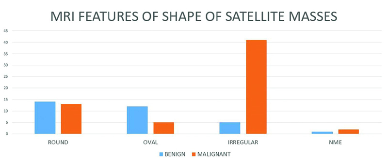
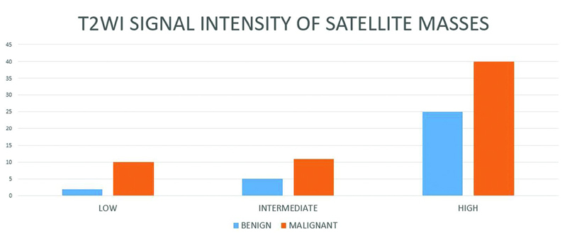
Sara Rehman The purpose of this study was to determine the diagnostic accuracy of breast magnetic resonance imaging (MRI) in classifying incidental satellite masses in biopsy-proven breast cancer patients as benign or malignant masses and assessing its impact on surgical management of these patients. We also analyzed the incidence of MRI-detected lesions, which were thereafter assessed with second look ultrasound (US). A retrospective study was performed on breast cancer patients presenting from August 01, 2016 to July 31, 2019, with satellite masses seen on base line MRI. Satellite masses were classified as benign and malignant based on MRI features of shape, margin, T2-weighted imaging signals, internal enhancement pattern, enhancement kinetic curves, and diffusion restriction. This was compared with results of histopathological examination. The number of MRI-detected lesions, location of the satellite mass, and type of surgery were also documented. Out of 400 breast cancer patients undergoing MRI breast, 115 patients had multiple masses. Histopathological diagnosis was available for 73 patients; and a total of 93 satellite masses were evaluated. There was evidence of additional masses on second look ultrasound in 21 patients. Of 72 masses classified as malignant on MRI, 58 showed malignant pathological outcome; while out of 21 masses characterized as benign on MRI, 18 turned out to be benign on histopathology. A statistically significant association was found between MRI features and pathological outcome of satellite masses ( = 0.001). The sensitivity, specificity, positive and negative predictive values, and accuracy were 95%, 56%, 80.56%, 85.7% and 81.7%, respectively. Based on these findings, modified radical mastectomy (MRM)/mastectomy was done for 42 patients, 5 patients underwent lumpectomy limited to a single tumor, extended resection done for 14 patients, 5 underwent bilateral breast conservation surgery (BCS), BCS for contralateral breast done for 4 patients undergoing ipsilateral MRM/mastectomy, and bilateral MRM/mastectomies were performed for 2 patients. One patient was lost to follow up. Breast MRI is the most sensitive modality for the assessment of breast cancer and plays an essential role in the detection of additional tumor foci. These findings can modify the surgical approach in these patients. However, considering the low specificity, biopsy of satellite masses is imperative to determine the most appropriate surgical plan.
萨拉·雷曼 本研究的目的是确定乳腺磁共振成像(MRI)在将活检证实的乳腺癌患者中的偶然卫星灶肿块分类为良性或恶性肿块方面的诊断准确性,并评估其对这些患者手术管理的影响。我们还分析了MRI检测到的病变的发生率,随后用二次超声(US)对其进行评估。 对2016年8月1日至2019年7月31日期间出现卫星灶肿块且基线MRI可见的乳腺癌患者进行了一项回顾性研究。根据形状、边缘、T2加权成像信号、内部强化模式、强化动力学曲线和扩散受限等MRI特征,将卫星灶肿块分为良性和恶性。将其与组织病理学检查结果进行比较。还记录了MRI检测到的病变数量、卫星灶肿块的位置和手术类型。 在400例接受乳腺MRI检查的乳腺癌患者中,115例患者有多个肿块。73例患者有组织病理学诊断;共评估了93个卫星灶肿块。21例患者在二次超声检查中有额外肿块的证据。在MRI上分类为恶性的72个肿块中,58个显示为恶性病理结果;而在MRI上特征为良性的21个肿块中,18个在组织病理学上被证明是良性的。发现MRI特征与卫星灶肿块的病理结果之间存在统计学显著关联(=0.001)。敏感性、特异性、阳性和阴性预测值以及准确性分别为95%、56%、80.56%、85.7%和81.7%。基于这些发现,42例患者进行了改良根治性乳房切除术(MRM)/乳房切除术,5例患者接受了仅限于单个肿瘤的肿块切除术,14例患者进行了扩大切除术,5例患者接受了双侧保乳手术(BCS),4例接受同侧MRM/乳房切除术的患者对侧乳房进行了BCS,2例患者进行了双侧MRM/乳房切除术。1例患者失访。 乳腺MRI是评估乳腺癌最敏感的方法,在检测额外肿瘤灶方面起着至关重要的作用。这些发现可以改变这些患者的手术方法。然而,考虑到特异性较低,对卫星灶肿块进行活检对于确定最合适的手术方案至关重要。