• 文献检索
  • 文档翻译
  • 深度研究
  • 学术资讯
  • Suppr Zotero 插件Zotero 插件
  • 邀请有礼
  • 套餐&价格
  • 历史记录
应用&插件
Suppr Zotero 插件Zotero 插件浏览器插件Mac 客户端Windows 客户端微信小程序
定价
高级版会员购买积分包购买API积分包
服务
文献检索文档翻译深度研究API 文档MCP 服务
关于我们
关于 Suppr公司介绍联系我们用户协议隐私条款
关注我们

Suppr 超能文献

核心技术专利:CN118964589B侵权必究
粤ICP备2023148730 号-1Suppr @ 2026

文献检索

告别复杂PubMed语法,用中文像聊天一样搜索,搜遍4000万医学文献。AI智能推荐,让科研检索更轻松。

立即免费搜索

文件翻译

保留排版,准确专业,支持PDF/Word/PPT等文件格式,支持 12+语言互译。

免费翻译文档

深度研究

AI帮你快速写综述,25分钟生成高质量综述,智能提取关键信息,辅助科研写作。

立即免费体验

未能通过产前诊断诊断出软骨发育不全症:一例报告。

Failure to diagnose hypochondroplasia by prenatal diagnosis: a case report.

机构信息

Department of Pediatrics, West China Second University Hospital, Sichuan University, Chengdu, China.

Ministry of Education Key Laboratory of Birth Defects and Related Diseases of Women and Children, Sichuan University, Chengdu, China.

出版信息

BMC Pediatr. 2023 Mar 2;23(1):100. doi: 10.1186/s12887-023-03917-2.

DOI:10.1186/s12887-023-03917-2
PMID:36859260
原文链接:https://pmc.ncbi.nlm.nih.gov/articles/PMC9979515/
Abstract

BACKGROUND

Hypochondroplasia (HCH) is a common nonlethal skeletal dysplasia caused by pathogenic variations in the fibroblast growth factor receptor 3 (FGFR3) gene, and HCH has similar clinical manifestations with achondroplasia (ACH), which can be screened during the fetal period by prenatal ultrasound testing and diagnosed by genetic testing.

CASE PRESENTATION

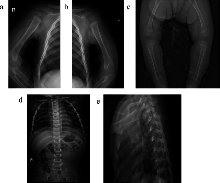
we report the special case of a patient with obvious growth retardation and rhizomelic disproportionate short stature, accompanied by other manifestations, including an enlarged head and short hands at 1 year old. However, several multiple color ultrasound exams identified shortened limbs (< 3rd percentile), an increased biparietal diameter (> 95th percentile) and a low nasal bridge in the fetal period. Due to the high incidence rate of ACH, genetic testing for the hotspot FGFR3 gene c.1138 g > A pathogenic variations was performed immediately in the third trimester. Unfortunately, the definitive diagnosis could not be made before birth due to the negative result of hotspot gene exam. Whole exome sequencing (WES) was performed at 1 year identified FGFR3 gene c.1620C > A variations positivity, and the patient was finally diagnosed as HCH.

CONCLUSION

Our report extends the understanding of the limitations of prenatal genetic diagnostic testing, especially the hot spot pathogenic variations test should be not the only clinical diagnostic basis. Moreover, this case also emphasizes that further gene analysis for patients with significant conflict between the clinical manifestation and the prenatal genetic panel examination findings should be reconducted timely to spare the family from a delayed diagnosis or a misdiagnosis.

摘要

背景

软骨发育不全症(HCH)是一种常见的非致死性骨骼发育不良疾病,由成纤维细胞生长因子受体 3(FGFR3)基因的致病变异引起,HCH 与软骨发育不全症(ACH)具有相似的临床表现,可以通过产前超声检查在胎儿期进行筛查,并通过基因检测进行诊断。

病例介绍

我们报告了一名患者的特殊病例,该患者存在明显的生长迟缓、四肢骨干短、四肢不成比例,伴有其他表现,包括头部增大和手部短。然而,几次多次彩色超声检查在胎儿期识别出四肢缩短(<第 3 百分位)、双额直径增加(>第 95 百分位)和鼻梁低。由于 ACH 的发病率高,立即在妊娠晚期对热点 FGFR3 基因 c.1138g> A 致病变异进行了基因检测。不幸的是,由于热点基因检测结果为阴性,出生前无法做出明确诊断。在 1 岁时进行全外显子组测序(WES)发现 FGFR3 基因 c.1620C> A 变异阳性,最终诊断为 HCH。

结论

我们的报告扩展了对产前遗传诊断检测局限性的认识,特别是热点致病变异检测不应是唯一的临床诊断依据。此外,该病例还强调,对于临床表现与产前基因panel 检查结果明显冲突的患者,应及时重新进行基因分析,以免家庭面临诊断延迟或误诊。

https://cdn.ncbi.nlm.nih.gov/pmc/blobs/e05b/9979515/908187380e12/12887_2023_3917_Fig3_HTML.jpg
https://cdn.ncbi.nlm.nih.gov/pmc/blobs/e05b/9979515/1cbd67796333/12887_2023_3917_Fig1_HTML.jpg
https://cdn.ncbi.nlm.nih.gov/pmc/blobs/e05b/9979515/af1c0d760dd6/12887_2023_3917_Fig2_HTML.jpg
https://cdn.ncbi.nlm.nih.gov/pmc/blobs/e05b/9979515/908187380e12/12887_2023_3917_Fig3_HTML.jpg
https://cdn.ncbi.nlm.nih.gov/pmc/blobs/e05b/9979515/1cbd67796333/12887_2023_3917_Fig1_HTML.jpg
https://cdn.ncbi.nlm.nih.gov/pmc/blobs/e05b/9979515/af1c0d760dd6/12887_2023_3917_Fig2_HTML.jpg
https://cdn.ncbi.nlm.nih.gov/pmc/blobs/e05b/9979515/908187380e12/12887_2023_3917_Fig3_HTML.jpg

相似文献

1
Failure to diagnose hypochondroplasia by prenatal diagnosis: a case report.未能通过产前诊断诊断出软骨发育不全症:一例报告。
BMC Pediatr. 2023 Mar 2;23(1):100. doi: 10.1186/s12887-023-03917-2.
2
Detection of a de novo Y278C mutation in FGFR3 in a pregnancy with severe fetal hypochondroplasia: prenatal diagnosis and literature review.一例患有严重胎儿软骨发育不全的妊娠中FGFR3基因新发Y278C突变的检测:产前诊断及文献综述
Taiwan J Obstet Gynecol. 2013 Dec;52(4):580-5. doi: 10.1016/j.tjog.2013.10.023.
3
Radiographic and genetic diagnosis of sporadic hypochondroplasia early in the neonatal period.新生儿期早期散发性软骨发育不全的影像学和遗传学诊断
Prenat Diagn. 2004 Jan;24(1):45-9. doi: 10.1002/pd.746.
4
Clinical and radiologic evaluation of a Turkish family with hypochondroplasia and a rare variant.土耳其一个伴有软骨发育不全症和罕见变异的家系的临床和影像学评估。
J Pediatr Endocrinol Metab. 2022 Apr 20;35(8):1097-1101. doi: 10.1515/jpem-2021-0773. Print 2022 Aug 26.
5
Achondroplasia-hypochondroplasia complex in a newborn infant.一名新生儿的软骨发育不全-软骨发育低下综合征
Am J Med Genet. 1999 Jun 11;84(5):396-400.
6
Prenatal diagnosis of hypochondroplasia: report of two cases.软骨发育不全的产前诊断:两例报告。
Am J Med Genet A. 2006 May 1;140(9):998-1003. doi: 10.1002/ajmg.a.31177.
7
Earlier detection of hypochondroplasia: A large single-center UK case series and systematic review.早发性软骨发育不全症的早期检测:一项大型英国单中心病例系列研究和系统评价。
Am J Med Genet A. 2021 Jan;185(1):73-82. doi: 10.1002/ajmg.a.61912. Epub 2020 Oct 14.
8
Compound heterozygosity for the Achondroplasia-hypochondroplasia FGFR3 mutations: prenatal diagnosis and postnatal outcome.软骨发育不全-软骨发育低下FGFR3基因突变的复合杂合性:产前诊断与产后结局
Am J Med Genet. 1999 Jun 11;84(5):401-5.
9
p.Ser348Cys mutation in FGFR3 gene leads to "Mild ACH /Severe HCH" phenotype.FGFR3基因中的p.Ser348Cys突变导致“轻度软骨发育不全/重度遗传性软骨发育不全”表型。
Eur J Med Genet. 2020 Feb;63(2):103659. doi: 10.1016/j.ejmg.2019.04.016. Epub 2019 Apr 30.
10
Identification of a novel mutation of FGFR3 gene in a large Chinese pedigree with hypochondroplasia by next-generation sequencing: A case report and brief literature review.通过二代测序技术在中国一个患有软骨发育不全的大型家系中鉴定FGFR3基因的新型突变:病例报告及文献综述
Medicine (Baltimore). 2019 Jan;98(4):e14157. doi: 10.1097/MD.0000000000014157.

本文引用的文献

1
Growth in achondroplasia including stature, weight, weight-for-height and head circumference from CLARITY: achondroplasia natural history study-a multi-center retrospective cohort study of achondroplasia in the US.成骨不全症的生长情况,包括身高、体重、体重身高比和头围:CLARITY:成骨不全症自然史研究——美国多中心回顾性队列研究成骨不全症。
Orphanet J Rare Dis. 2021 Dec 23;16(1):522. doi: 10.1186/s13023-021-02141-4.
2
International Consensus Statement on the diagnosis, multidisciplinary management and lifelong care of individuals with achondroplasia.关于软骨发育不全个体的诊断、多学科管理及终身护理的国际共识声明
Nat Rev Endocrinol. 2022 Mar;18(3):173-189. doi: 10.1038/s41574-021-00595-x. Epub 2021 Nov 26.
3
Diagnostic, treatment and outcome possibilities in achondroplasia.
软骨发育不全的诊断、治疗及预后可能性
Med Pharm Rep. 2021 Aug;94(Suppl No 1):S22-S24. doi: 10.15386/mpr-2222. Epub 2021 Aug 10.
4
[Experts consensus on diagnosis and treatment of achondroplasia].[软骨发育不全诊断与治疗专家共识]
Zhonghua Er Ke Za Zhi. 2021 Jul 2;59(7):545-550. doi: 10.3760/cma.j.cn112140-20201229-01142.
5
The role of sonographic phenotyping in delivering an efficient noninvasive prenatal diagnosis service for FGFR3-related skeletal dysplasias.超声表型分析在提供 FGFR3 相关骨骼发育不良的高效无创性产前诊断服务中的作用。
Prenat Diagn. 2020 Jun;40(7):785-791. doi: 10.1002/pd.5687. Epub 2020 May 25.
6
A framework for the radiologic diagnosis of skeletal dysplasias and syndromes as revealed by molecular genetics.基于分子遗传学揭示的骨骼发育不良和综合征的放射学诊断框架。
Pediatr Radiol. 2019 Nov;49(12):1576-1586. doi: 10.1007/s00247-019-04545-8. Epub 2019 Nov 4.
7
A sonographic approach to the prenatal diagnosis of skeletal dysplasias.超声在骨骼发育不良产前诊断中的应用。
Prenat Diagn. 2019 Aug;39(9):701-719. doi: 10.1002/pd.5501. Epub 2019 Jul 14.
8
Molecular mechanisms of fibroblast growth factor signaling in physiology and pathology.成纤维细胞生长因子信号在生理和病理中的分子机制。
Cold Spring Harb Perspect Biol. 2013 Jun 1;5(6):a015958. doi: 10.1101/cshperspect.a015958.
9
Prenatal diagnosis of skeletal dysplasia due to FGFR3 gene mutations: a 9-year experience : prenatal diagnosis in FGFR3 gene.FGFR3 基因突变所致骨骼发育不良的产前诊断:9 年经验总结:FGFR3 基因的产前诊断。
J Assist Reprod Genet. 2009 Aug;26(8):455-60. doi: 10.1007/s10815-009-9339-1. Epub 2009 Sep 30.
10
Prenatal sonographic diagnosis of skeletal dysplasias.产前超声诊断骨骼发育不良。
Ultrasound Obstet Gynecol. 2009 Aug;34(2):160-70. doi: 10.1002/uog.6359.