Njei Basile, McCarty Thomas R, Mohan Babu P, Fozo Lydia, Navaneethan Udayakumar
Global Clinical Scholars Program, Harvard Medical School, Boston, MA, USA (Basile Njei).
Investigative Medicine Program, Yale University School of Medicine, New Haven, CT, USA (Basile Njei).
Ann Gastroenterol. 2023 Mar-Apr;36(2):223-230. doi: 10.20524/aog.2023.0779. Epub 2023 Feb 2.
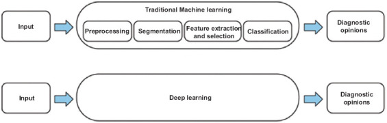
Artificial intelligence (AI), when applied to computer vision using a convolutional neural network (CNN), is a promising tool in "difficult-to-diagnose" conditions such as malignant biliary strictures and cholangiocarcinoma (CCA). The aim of this systematic review is to summarize and review the available data on the diagnostic utility of endoscopic AI-based imaging for malignant biliary strictures and CCA.
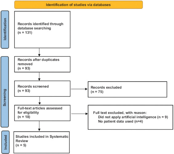
In this systematic review, PubMed, Scopus and Web of Science databases were reviewed for studies published from January 2000 to June 2022. Extracted data included type of endoscopic imaging modality, AI classifiers, and performance measures.
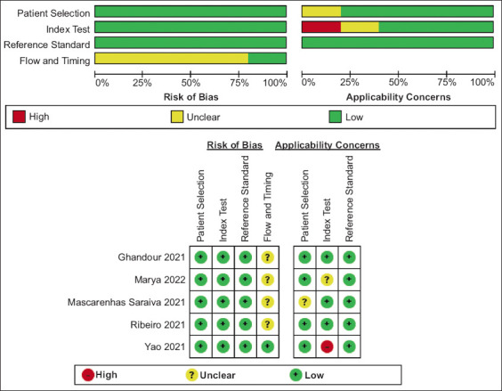
The search yielded 5 studies involving 1465 patients. Of the 5 included studies, 4 (n=934; 3,775,819 images) used CNN in combination with cholangioscopy, while one study (n=531; 13,210 images) used CNN with endoscopic ultrasound (EUS). The average image processing speed of CNN with cholangioscopy was 7-15 msec per frame while that of CNN with EUS was 200-300 msec per frame. The highest performance metrics were observed with CNN-cholangioscopy (accuracy 94.9%, sensitivity 94.7%, and specificity 92.1%). CNN-EUS was associated with the greatest clinical performance application, providing station recognition and bile duct segmentation; thus reducing procedure length and providing real-time feedback to the endoscopist.
Our results suggest that there is increasing evidence to support a role for AI in the diagnosis of malignant biliary strictures and CCA. CNN-based machine leaning of cholangioscopy images appears to be the most promising, while CNN-EUS has the best clinical performance application.
人工智能(AI)应用于使用卷积神经网络(CNN)的计算机视觉时,在恶性胆管狭窄和胆管癌(CCA)等“难以诊断”的病症中是一种很有前景的工具。本系统评价的目的是总结和回顾关于基于内镜AI成像对恶性胆管狭窄和CCA的诊断效用的现有数据。
在本系统评价中,检索了PubMed、Scopus和Web of Science数据库中2000年1月至2022年6月发表的研究。提取的数据包括内镜成像方式的类型、AI分类器和性能指标。
检索得到5项研究,涉及1465例患者。在纳入的5项研究中,4项(n = 934;3,775,819张图像)将CNN与胆管镜检查相结合,而1项研究(n = 531;13,210张图像)将CNN与内镜超声(EUS)结合使用。CNN与胆管镜检查结合时的平均图像处理速度为每帧7 - 15毫秒,而CNN与EUS结合时为每帧200 - 300毫秒。CNN - 胆管镜检查的性能指标最高(准确率94.9%,灵敏度94.7%,特异性92.1%)。CNN - EUS具有最大的临床性能应用,可提供部位识别和胆管分割;从而缩短手术时间并为内镜医师提供实时反馈。
我们的结果表明,越来越多的证据支持AI在恶性胆管狭窄和CCA诊断中的作用。基于CNN的胆管镜图像机器学习似乎最有前景,而CNN - EUS具有最佳的临床性能应用。