Rayisi Mahdiye, Malekzadeh Kianoosh, Afsa Masoomeh
Dept. of Maxillofacial Radiology, Faculty of Dentistry, Hormozgan University of Medical Sciences, Bandar Abbas, Iran.
Hormozgan Health Institute, Hormozgan University of Medical Sciences, Bandar Abbas, Iran.
J Dent (Shiraz). 2023 Mar;24(1):7-11. doi: 10.30476/DENTJODS.2021.92400.1638.
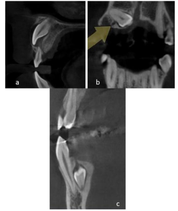
Gubernacular canal (GC) is a canal that extends from the follicle of unerupted permanent teeth to the alveolar bone crest filled with remnants of the dental lamina. This canal is thought to guide tooth eruption and be related to some pathologic conditions.
This study aimed to determine the presence of GC and its anatomical characteristics in teeth, which failed to erupt normally on cone beam computed tomography (CBCT) images.
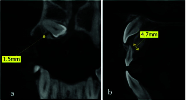
This cross-sectional study evaluated CBCT images of 77 impacted permanent and supernumerary teeth obtained from 29 females and 21 males. The frequency of GC detection, its location in relation to the crown and root, the anatomical surface of the tooth from which the canal has originated, and the adjacent cortical table to which the canal opens, along with the length of the GC were studied.
GC was observed in 53.2% of teeth. The anatomical tooth aspect of origin was occlusal/ incisal in 41.5% and crown in 82.9% of teeth. Moreover, 51.2% of GCs opened in palatal/lingual cortex and 63.4% of canals were not located along the tooth long axis. Finally, GC was detected in 85.7% of teeth undergoing the crown formation stage.
Although GC was introduced as an eruption pathway, this canal is also present in impacted teeth. This means that presence of this canal does not promise the normal eruption of tooth and the anatomical characteristics of GC may influence the eruption process.
gubernacular管(GC)是一条从未萌出恒牙的牙囊延伸至牙槽嵴顶的管道,内充满牙板遗迹。该管道被认为引导牙齿萌出,并与一些病理状况相关。
本研究旨在确定在锥形束计算机断层扫描(CBCT)图像上未能正常萌出的牙齿中GC的存在及其解剖特征。
本横断面研究评估了从29名女性和21名男性获取的77颗阻生恒牙和多生牙的CBCT图像。研究了GC的检出频率、其相对于牙冠和牙根的位置、管道起源的牙齿解剖面、管道开口的相邻皮质骨板以及GC的长度。
在53.2%的牙齿中观察到GC。管道起源的牙齿解剖面在41.5%的牙齿中为咬合面/切缘,在82.9%的牙齿中为牙冠。此外,51.2%的GC开口于腭侧/舌侧皮质骨板,63.4%的管道不沿牙齿长轴走行。最后,在85.7%处于牙冠形成阶段的牙齿中检测到GC。
尽管GC被认为是一条萌出途径,但该管道也存在于阻生牙中。这意味着该管道的存在并不能保证牙齿正常萌出,且GC的解剖特征可能影响萌出过程。