Department of Molecular and Integrative Physiology, University of Illinois at Urbana-Champaign, Urbana, IL, 61801, USA.
Department of Animal Sciences, University of Illinois at Urbana-Champaign, 1207 W. Gregory Drive, Room 314 ASL, Urbana, IL, 61801, USA.
J Neuroinflammation. 2023 Mar 6;20(1):59. doi: 10.1186/s12974-023-02713-0.
Chronic pelvic pain (CPP) is a common symptom of endometriosis. Women with endometriosis are also at a high risk of suffering from anxiety, depression, and other psychological disorders. Recent studies indicate that endometriosis can affect the central nervous system (CNS). Changes in the functional activity of neurons, functional magnetic resonance imaging signals, and gene expression have been reported in the brains of rat and mouse models of endometriosis. The majority of the studies thus far have focused on neuronal changes, whereas changes in the glial cells in different brain regions have not been studied.
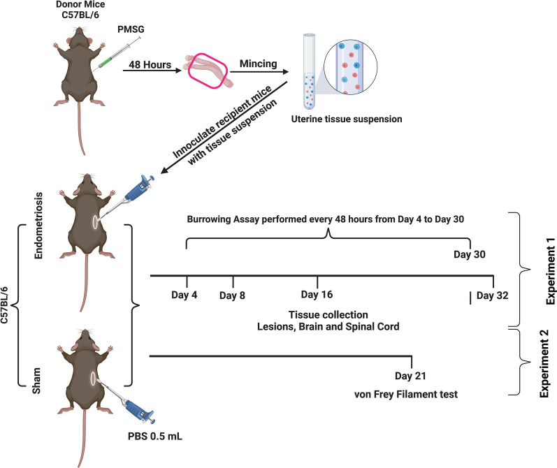
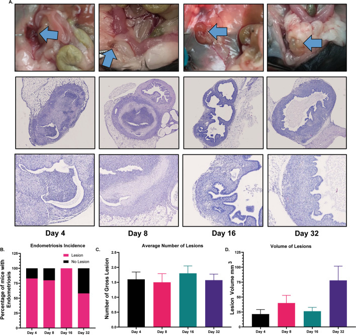
Endometriosis was induced in female mice (45-day-old; n = 6-11/timepoint) by syngeneic transfer of donor uterine tissue into the peritoneal cavity of recipient animals. Brains, spines, and endometriotic lesions were collected for analysis at 4, 8, 16, and 32 days post-induction. Sham surgery mice were used as controls (n = 6/timepoint). The pain was assessed using behavioral tests. Using immunohistochemistry for microglia marker ionized calcium-binding adapter molecule-1 (IBA1) and machine learning "Weka trainable segmentation" plugin in Fiji, we evaluated the morphological changes in microglia in different brain regions. Changes in glial fibrillary acidic protein (GFAP) for astrocytes, tumor necrosis factor (TNF), and interleukin-6 (IL6) were also evaluated.
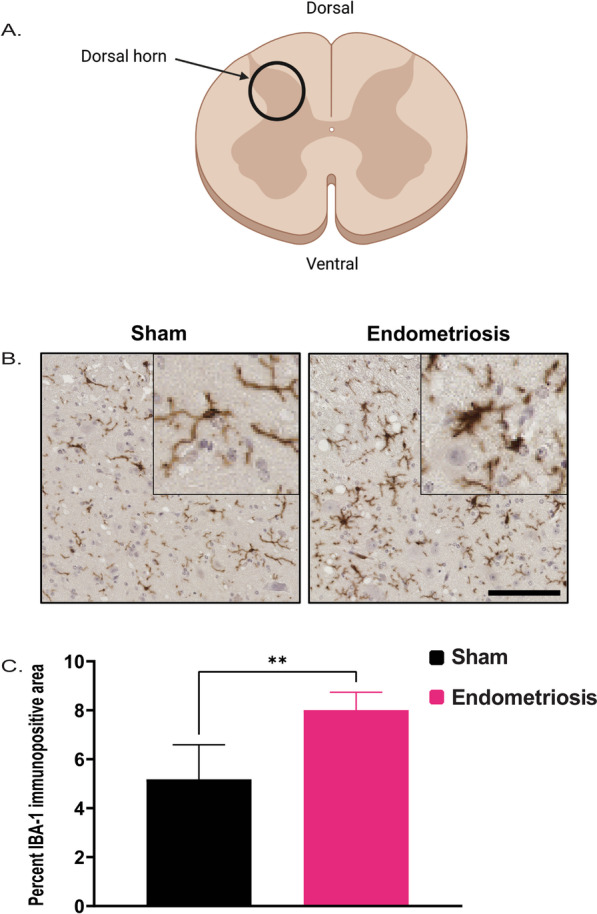
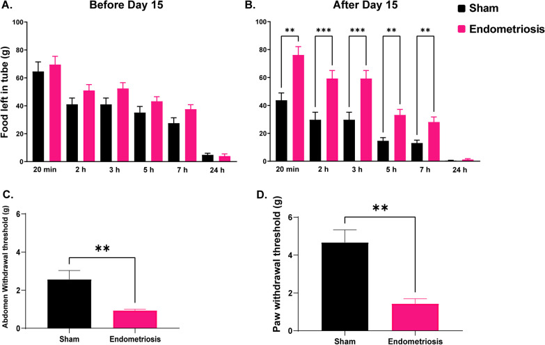
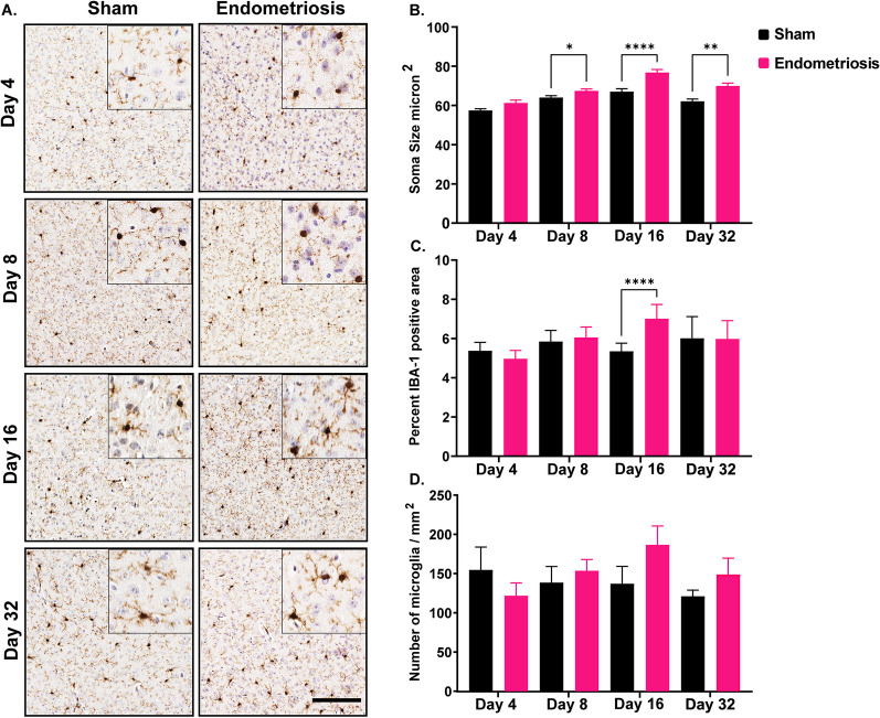
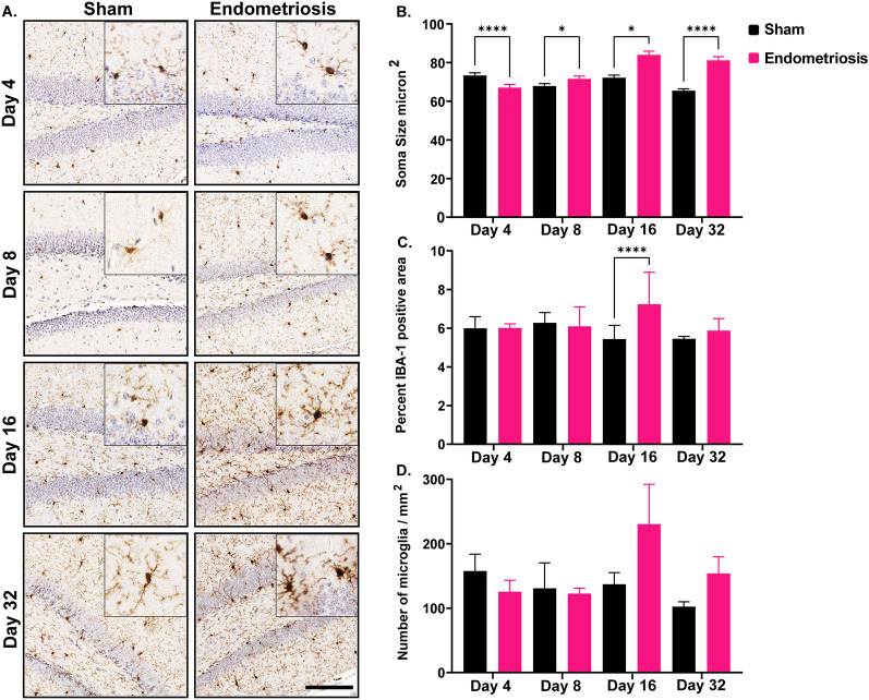
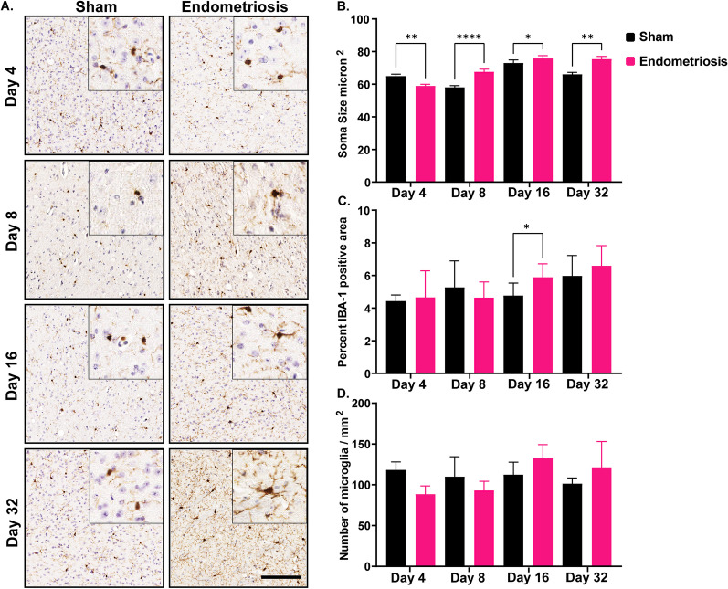
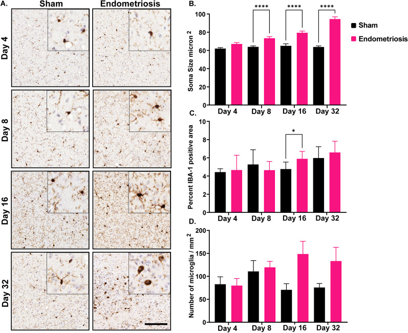
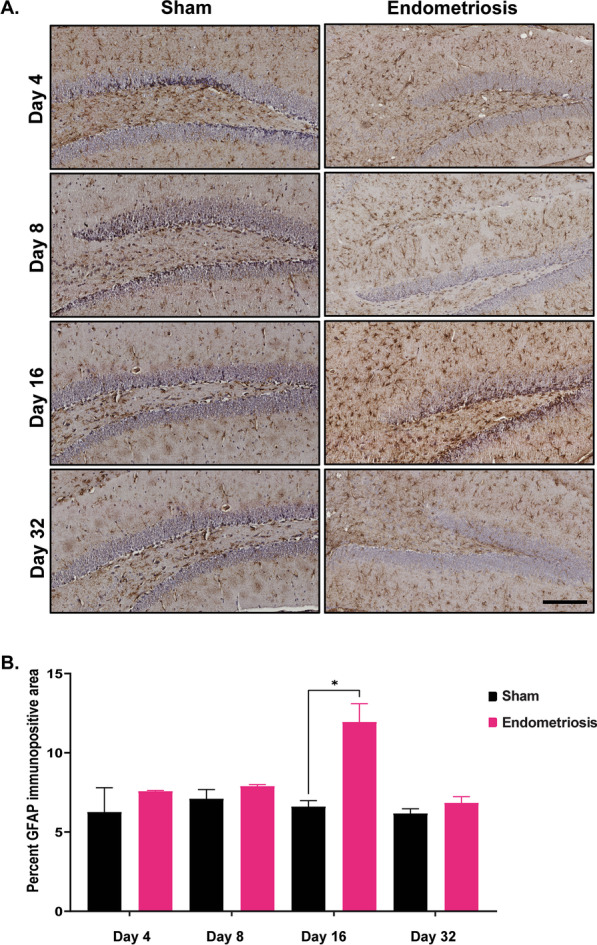
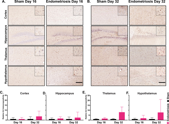
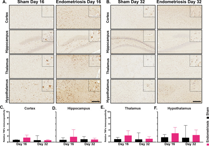
We observed an increase in microglial soma size in the cortex, hippocampus, thalamus, and hypothalamus of mice with endometriosis compared to sham controls on days 8, 16, and 32. The percentage of IBA1 and GFAP-positive area was increased in the cortex, hippocampus, thalamus, and hypothalamus in mice with endometriosis compared to sham controls on day 16. The number of microglia and astrocytes did not differ between endometriosis and sham control groups. We observed increased TNF and IL6 expression when expression levels from all brain regions were combined. Mice with endometriosis displayed reduced burrowing behavior and hyperalgesia in the abdomen and hind-paw.
We believe this is the first report of central nervous system-wide glial activation in a mouse model of endometriosis. These results have significant implications for understanding chronic pain associated with endometriosis and other issues such as anxiety and depression in women with endometriosis.
慢性盆腔疼痛(CPP)是子宫内膜异位症的常见症状。患有子宫内膜异位症的女性也有很高的患焦虑、抑郁和其他心理障碍的风险。最近的研究表明,子宫内膜异位症会影响中枢神经系统(CNS)。在子宫内膜异位症的大鼠和小鼠模型的大脑中,已经报道了神经元功能活动的变化、功能磁共振成像信号和基因表达的变化。迄今为止,大多数研究都集中在神经元变化上,而不同脑区的神经胶质细胞变化尚未得到研究。
通过将供体子宫组织同种异体移植到受体动物的腹腔中,在雌性小鼠(45 天大;n = 6-11/时间点)中诱导子宫内膜异位症。在诱导后 4、8、16 和 32 天收集大脑、脊柱和子宫内膜异位病灶进行分析。假手术小鼠作为对照(n = 6/时间点)。使用行为测试评估疼痛。使用免疫组织化学检测小胶质细胞标志物离子钙结合接头蛋白-1(IBA1)和 Fiji 中的机器学习“Weka 可训练分割”插件,我们评估了不同脑区小胶质细胞形态的变化。还评估了星形胶质细胞的胶质纤维酸性蛋白(GFAP)、肿瘤坏死因子(TNF)和白细胞介素 6(IL6)的变化。
与假手术对照组相比,我们观察到在诱导后 8、16 和 32 天,子宫内膜异位症小鼠的大脑皮层、海马体、丘脑和下丘脑的小胶质细胞体大小增加。与假手术对照组相比,在诱导后 16 天,子宫内膜异位症小鼠的大脑皮层、海马体、丘脑和下丘脑的 IBA1 和 GFAP 阳性面积百分比增加。子宫内膜异位症组和假手术对照组的小胶质细胞和星形胶质细胞数量没有差异。当结合所有脑区的表达水平时,我们观察到 TNF 和 IL6 表达增加。患有子宫内膜异位症的小鼠表现出腹部和后足的挖掘行为减少和痛觉过敏。
我们相信这是子宫内膜异位症小鼠模型中中枢神经系统广泛神经胶质激活的首次报道。这些结果对理解与子宫内膜异位症相关的慢性疼痛以及子宫内膜异位症女性的焦虑和抑郁等问题具有重要意义。