Vanderbilt University Institute of Imaging Science, Vanderbilt University Medical Center, Nashville, TN, USA; Department of Radiology and Radiological Sciences, Vanderbilt University Medical Center, Nashville, TN, USA.
Department of Radiology and Radiological Sciences, Vanderbilt University Medical Center, Nashville, TN, USA; Department of Neurology, Vanderbilt University Medical Center, Nashville, TN, USA.
Neuroimage Clin. 2023;37:103366. doi: 10.1016/j.nicl.2023.103366. Epub 2023 Mar 2.
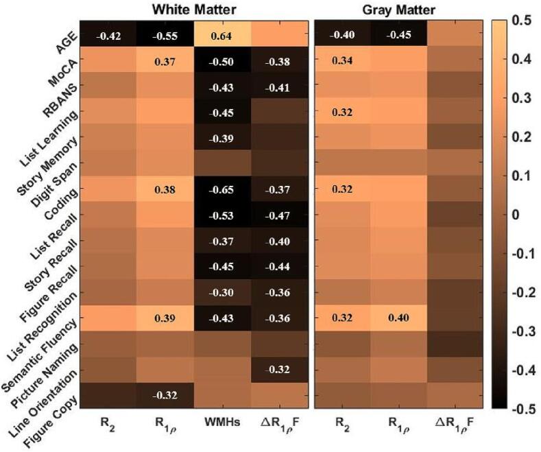
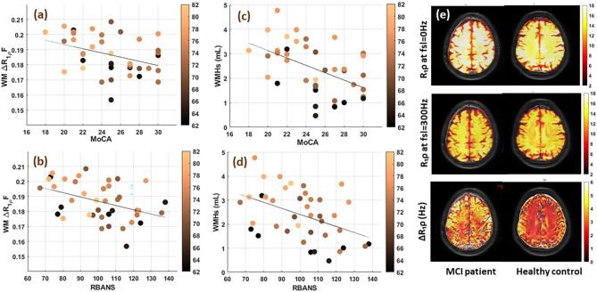
Much previous neuroimaging research in Alzheimers disease has focused on the roles of amyloid and tau proteins, but recent studies have implicated microvascular changes in white matter as early indicators of damage related to later dementia. We used MRI to derive novel, non-invasive measurements of R dispersion using different locking fields to characterize variations of microvascular structure and integrity in brain tissues. We developed a non-invasive 3D R dispersion imaging technique using different locking fields at 3T. We acquired MR images and cognitive assessments of participants with mild cognitive impairment (MCI) and compared them to age-matched healthy controls in a cross-sectional study. After providing informed consent, 40 adults aged 62 to 82 years (n = 17 MCI) were included in this study. White matter ΔR-fraction measured by R dispersion imaging showed a strong correlation with the cognitive status of older adults (β = -0.4, p-value < 0.01) independent of age, in contrast to other conventional MRI markers such as T, R, and white matter hyperintense lesion volume (WMHs) measured with T-FLAIR. The correlation of WMHs with cognitive status was no longer significant after adjusting for age and sex in linear regression analysis, and the size of the regression coefficient was substantially decreased (53% lower). This work establishes a new non-invasive method that potentially characterizes impairment of the microvascular structure of white matter in MCI patients compared to healthy controls. The application of this method in longitudinal studies would improve our fundamental understanding of the pathophysiologic changes that accompany abnormal cognitive decline with aging and help identify potential targets for treatment of Alzheimer's disease.
先前许多阿尔茨海默病的神经影像学研究都集中在淀粉样蛋白和tau 蛋白的作用上,但最近的研究表明,脑白质微血管的变化是与后期痴呆相关损害的早期指标。我们使用 MRI 技术,通过不同的锁定场获得了新的、非侵入性的 R 分散测量值,以表征脑组织中微血管结构和完整性的变化。我们在 3T 下开发了一种使用不同锁定场的非侵入性 3D R 分散成像技术。我们对轻度认知障碍 (MCI) 患者进行了磁共振成像和认知评估,并在一项横断面研究中与年龄匹配的健康对照组进行了比较。在提供知情同意后,共有 40 名年龄在 62 至 82 岁之间的成年人(n = 17 名 MCI)参与了这项研究。通过 R 分散成像测量的白质 ΔR-分数与老年人的认知状态密切相关(β = -0.4,p 值 < 0.01),与其他常规 MRI 标志物(如 T、R 和 T-FLAIR 测量的白质高信号病变体积 [WMHs])不同。在线性回归分析中,调整年龄和性别后,WMHs 与认知状态的相关性不再显著,回归系数的大小大大降低(降低 53%)。这项工作建立了一种新的非侵入性方法,该方法可能与健康对照组相比,能够对 MCI 患者脑白质微血管结构的损伤进行特征描述。该方法在纵向研究中的应用将提高我们对与衰老相关的异常认知下降伴随的病理生理变化的基本认识,并有助于确定治疗阿尔茨海默病的潜在靶点。