• 文献检索
  • 文档翻译
  • 深度研究
  • 学术资讯
  • Suppr Zotero 插件Zotero 插件
  • 邀请有礼
  • 套餐&价格
  • 历史记录
应用&插件
Suppr Zotero 插件Zotero 插件浏览器插件Mac 客户端Windows 客户端微信小程序
定价
高级版会员购买积分包购买API积分包
服务
文献检索文档翻译深度研究API 文档MCP 服务
关于我们
关于 Suppr公司介绍联系我们用户协议隐私条款
关注我们

Suppr 超能文献

核心技术专利:CN118964589B侵权必究
粤ICP备2023148730 号-1Suppr @ 2026

文献检索

告别复杂PubMed语法,用中文像聊天一样搜索,搜遍4000万医学文献。AI智能推荐,让科研检索更轻松。

立即免费搜索

文件翻译

保留排版,准确专业,支持PDF/Word/PPT等文件格式,支持 12+语言互译。

免费翻译文档

深度研究

AI帮你快速写综述,25分钟生成高质量综述,智能提取关键信息,辅助科研写作。

立即免费体验

心内超声引导下左束支近端起搏的植入。

Intracardiac Echocardiography-Guided Implantation for Proximal Left Bundle Branch Pacing.

机构信息

Division of Cardiology, Yunnan Arrhythmia Research Center, the First People's Hospital of Yunnan Province, the Affiliated Hospital of Kunming University of Science and Technology, China (X.K., X.Z., Y.C., F.W., P.W., X.G., H.X., H.W., L.-L.W., J.F.).

CRHF Division, Medtronic PLC, Mounds View, MN (X.Z.).

出版信息

Circ Arrhythm Electrophysiol. 2023 Apr;16(4):e011408. doi: 10.1161/CIRCEP.122.011408. Epub 2023 Mar 16.

DOI:10.1161/CIRCEP.122.011408
PMID:36924221
原文链接:https://pmc.ncbi.nlm.nih.gov/articles/PMC10108837/
Abstract

BACKGROUND

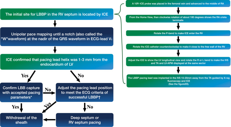
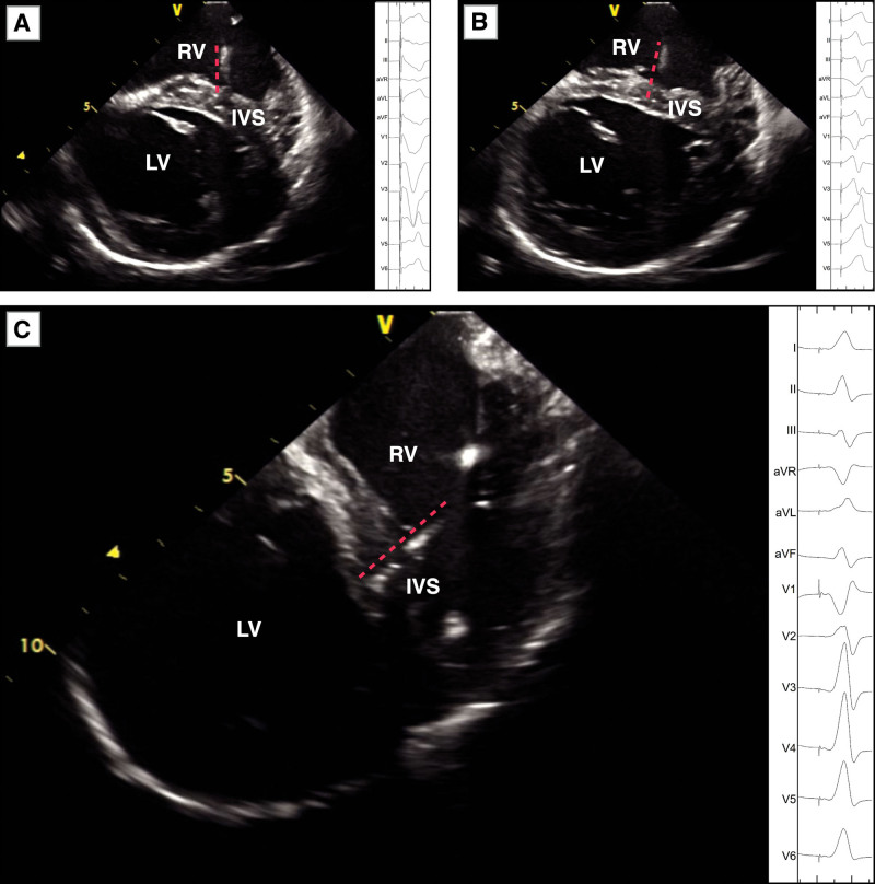
Multiple screw-in attempts under fluoroscopy are often needed to place the pacing lead tip near or at the left bundle branch (LBB). This study was conducted to evaluate the feasibility of implanting an LBB pacing lead in the proximal LBB (PLBB) guided by intracardiac echocardiography (ICE).

METHODS

The distribution of the LBB was initially determined by ICE anatomic imaging and 3-dimensional electrical mapping of His and LBB potentials in 20 patients in the first parts of the study. In the second part, 101 consecutive pacemaker-indicated patients were randomized into the ICE-guided and non-ICE groups for LBB pacing implantation. The procedural details and electrophysiological characteristics of the 2 groups were compared.

RESULTS

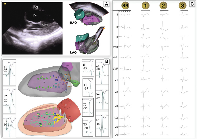
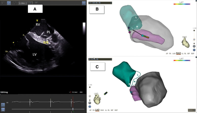
In the first part of the study, PLBB was identified at 10 to 20 mm from the tricuspid annulus toward the apex with an area of 4.5±1.1 cm. In the second part, the number of lead screw-in attempts in the septum was fewer in the ICE group than in the non-ICE group (1.43±0.62 versus 1.98±0.75, =0.0002). The duration of the procedure (26±8 versus 43±9 minutes, <0.001) and fluoroscopy for LBB pacing implantation (7.4±1.8 versus 10.7±2.4 minutes, <0.001) in the ICE group was significantly shorter than those in the non-ICE group. LBB pacing in the ICE group generated a lesser QRS duration with more cases of LBB trunk pacing (46.8% versus 25%, =0.031) and PLBB (91.5% versus 72.7%, =0.0267) pacing compared with that in the non-ICE group.

CONCLUSIONS

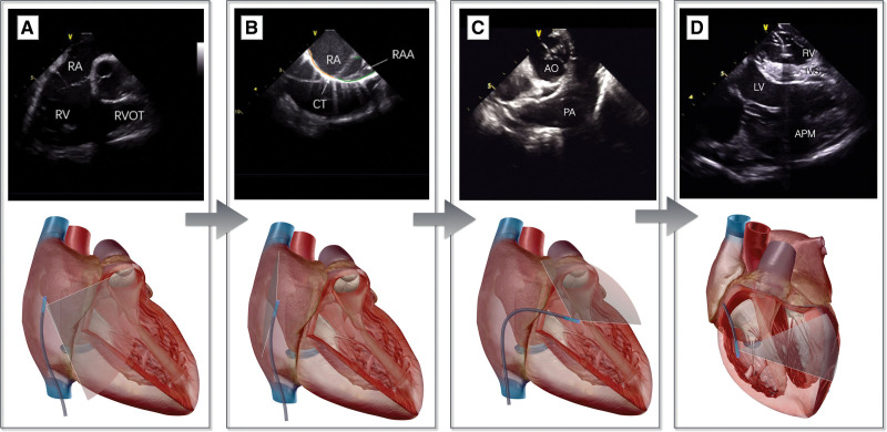
The basal left ventricular septum can be better visualized using ICE. ICE-guided PLBB pacing is feasible and safe, with a shorter duration required for the procedure and fluoroscopy, and generates greater LBB trunk pacing and PLBB pacing.

摘要

背景

在透视引导下,通常需要多次尝试将起搏导线尖端放置在左束支(LBB)附近或处。本研究旨在评估在心脏内超声心动图(ICE)引导下植入 LBB 起搏导线的可行性。

方法

在研究的第一部分,通过 ICE 解剖成像和希氏束和 LBB 电位的 3 维电描记图初步确定 LBB 的分布。在第二部分,将 101 例连续的起搏器指征患者随机分为 ICE 引导组和非 ICE 组进行 LBB 起搏植入。比较两组的手术细节和电生理特征。

结果

在研究的第一部分,在距离三尖瓣环 10 至 20mm 处向心尖方向的间隔内识别出 PLBB,面积为 4.5±1.1cm。在第二部分,ICE 组比非 ICE 组在间隔内导丝插入次数更少(1.43±0.62 比 1.98±0.75,=0.0002)。ICE 组的手术时间(26±8 比 43±9 分钟,<0.001)和 LBB 起搏植入的透视时间(7.4±1.8 比 10.7±2.4 分钟,<0.001)明显短于非 ICE 组。ICE 组的 LBB 起搏产生的 QRS 持续时间更短,并且 LBB 干起搏(46.8%比 25%,=0.031)和 PLBB(91.5%比 72.7%,=0.0267)起搏的病例更多。

结论

ICE 可更好地显示左心室间隔基底部。ICE 引导的 PLBB 起搏是可行且安全的,需要的手术和透视时间更短,产生更大的 LBB 干起搏和 PLBB 起搏。

https://cdn.ncbi.nlm.nih.gov/pmc/blobs/e1aa/10108837/33bdc4237076/hae-16-e011408-g006.jpg
https://cdn.ncbi.nlm.nih.gov/pmc/blobs/e1aa/10108837/f71d1a3772b5/hae-16-e011408-g001.jpg
https://cdn.ncbi.nlm.nih.gov/pmc/blobs/e1aa/10108837/c95c2e51f7c6/hae-16-e011408-g003.jpg
https://cdn.ncbi.nlm.nih.gov/pmc/blobs/e1aa/10108837/a747e8c6a632/hae-16-e011408-g004.jpg
https://cdn.ncbi.nlm.nih.gov/pmc/blobs/e1aa/10108837/2c5609a50476/hae-16-e011408-g005.jpg
https://cdn.ncbi.nlm.nih.gov/pmc/blobs/e1aa/10108837/33bdc4237076/hae-16-e011408-g006.jpg

相似文献

1
Intracardiac Echocardiography-Guided Implantation for Proximal Left Bundle Branch Pacing.心内超声引导下左束支近端起搏的植入。
Circ Arrhythm Electrophysiol. 2023 Apr;16(4):e011408. doi: 10.1161/CIRCEP.122.011408. Epub 2023 Mar 16.
2
Permanent left bundle branch area pacing utilizing intracardiac echocardiogram.利用心内超声心动图进行永久性左束支区域起搏。
BMC Cardiovasc Disord. 2020 Aug 18;20(1):377. doi: 10.1186/s12872-020-01649-0.
3
Left bundle branch potential predicts better electrical synchrony in bradycardia patients receiving left bundle branch pacing.左束支电位预测左束支起搏治疗心动过缓患者具有更好的电同步性。
BMC Cardiovasc Disord. 2022 Aug 19;22(1):376. doi: 10.1186/s12872-022-02812-5.
4
Prospective evaluation of feasibility and electrophysiologic and echocardiographic characteristics of left bundle branch area pacing.前瞻性评估左束支区域起搏的可行性及电生理和超声心动图特征。
Heart Rhythm. 2019 Dec;16(12):1774-1782. doi: 10.1016/j.hrthm.2019.05.011. Epub 2019 May 25.
5
Evaluation of the Criteria to Distinguish Left Bundle Branch Pacing From Left Ventricular Septal Pacing.评价区分左束支起搏与左心室间隔起搏的标准。
JACC Clin Electrophysiol. 2021 Sep;7(9):1166-1177. doi: 10.1016/j.jacep.2021.02.018. Epub 2021 Apr 28.
6
A novel 9-partition method using fluoroscopic images for guiding left bundle branch pacing.一种新的 9 分区方法,使用荧光透视图像引导左束支起搏。
Heart Rhythm. 2020 Oct;17(10):1759-1767. doi: 10.1016/j.hrthm.2020.05.018. Epub 2020 May 15.
7
Electrophysiological Insights into Three Modalities of Left Bundle Branch Area Pacing in Patients Indicated for Pacing Therapy.三种左束支区域起搏模式的电生理见解在需要起搏治疗的患者中。
Int Heart J. 2021 Jan 30;62(1):78-86. doi: 10.1536/ihj.20-490. Epub 2020 Dec 26.
8
Characteristics of intracardiac electrogram of the interventricular septum in the left bundle branch pacing.左束支起搏时心室内电图的特征。
BMC Cardiovasc Disord. 2022 Jun 17;22(1):274. doi: 10.1186/s12872-022-02715-5.
9
Feasibility and cardiac synchrony of permanent left bundle branch pacing through the interventricular septum.经室间隔行永久性左束支起搏的可行性和心脏同步性。
Europace. 2019 Nov 1;21(11):1694-1702. doi: 10.1093/europace/euz188.
10
Relationship of paced left bundle branch pacing morphology with anatomic location and physiological outcomes.起搏左束支区域形态与解剖位置和生理结果的关系。
Heart Rhythm. 2021 Jun;18(6):946-953. doi: 10.1016/j.hrthm.2021.03.034. Epub 2021 Mar 27.

引用本文的文献

1
A systematic review and meta-analysis of the impact of left bundle branch area pacing on right ventricular function.左束支区域起搏对右心室功能影响的系统评价与荟萃分析
Front Cardiovasc Med. 2025 Jan 30;12:1545757. doi: 10.3389/fcvm.2025.1545757. eCollection 2025.
2
Current Advance, Challenges and Future Perspectives of Conduction System Pacing.传导系统起搏的当前进展、挑战及未来展望
Rev Cardiovasc Med. 2024 Dec 16;25(12):438. doi: 10.31083/j.rcm2512438. eCollection 2024 Dec.
3
Left bundle branch area pacing guided by intracardiac echocardiography imaging.
心腔内超声心动图成像引导下的左束支区域起搏
Heart Rhythm O2. 2024 Aug 30;5(11):842-843. doi: 10.1016/j.hroo.2024.08.012. eCollection 2024 Nov.
4
Enhancing cardiac pacing strategies: a review of conduction system pacing compared with right and biventricular pacing and their influence on myocardial function.增强心脏起搏策略:比较心脏传导系统起搏与右心室和双心室起搏及其对心肌功能影响的综述。
Eur Heart J Cardiovasc Imaging. 2024 Jun 28;25(7):879-887. doi: 10.1093/ehjci/jeae090.
5
Intracardiac Echocardiography Guidance for Left Bundle Pacing: An Expensive Adjunct or Necessity for Optimal Lead Placement?心腔内超声心动图指导左束支起搏:是昂贵的辅助手段还是优化导线放置的必要条件?
Circ Arrhythm Electrophysiol. 2023 Apr;16(4):e011931. doi: 10.1161/CIRCEP.123.011931. Epub 2023 Mar 23.