Department of Gastroenterology, The First Affiliated Hospital, Zhejiang University School of Medicine, Hangzhou, 310003, Zhejiang, China.
BMC Infect Dis. 2023 Mar 21;23(1):172. doi: 10.1186/s12879-023-08095-1.
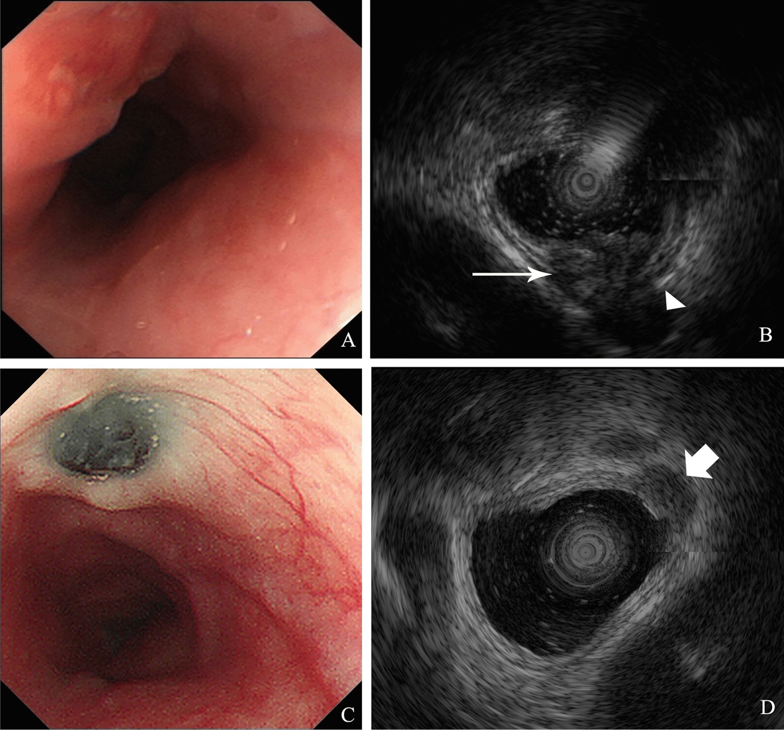
Anthracosis is a disease generally considered to be in the lungs resulting from exposure to industrial dust in the workplace. Esophageal anthracosis is a fairly rare phenomenon and shows a strong correlation with tuberculosis. Moreover, esophageal involvement in tuberculosis is also rare. We here present an extremely rare case in which follow-up gastroesophageal endoscopy revealed a mass with a sunken, black area in the center and raised ring-like pattern in the surrounding mucosa resembling malignant melanoma. Uncovering the patient's tuberculosis history finally avoided a misdiagnosis or overtreatment.
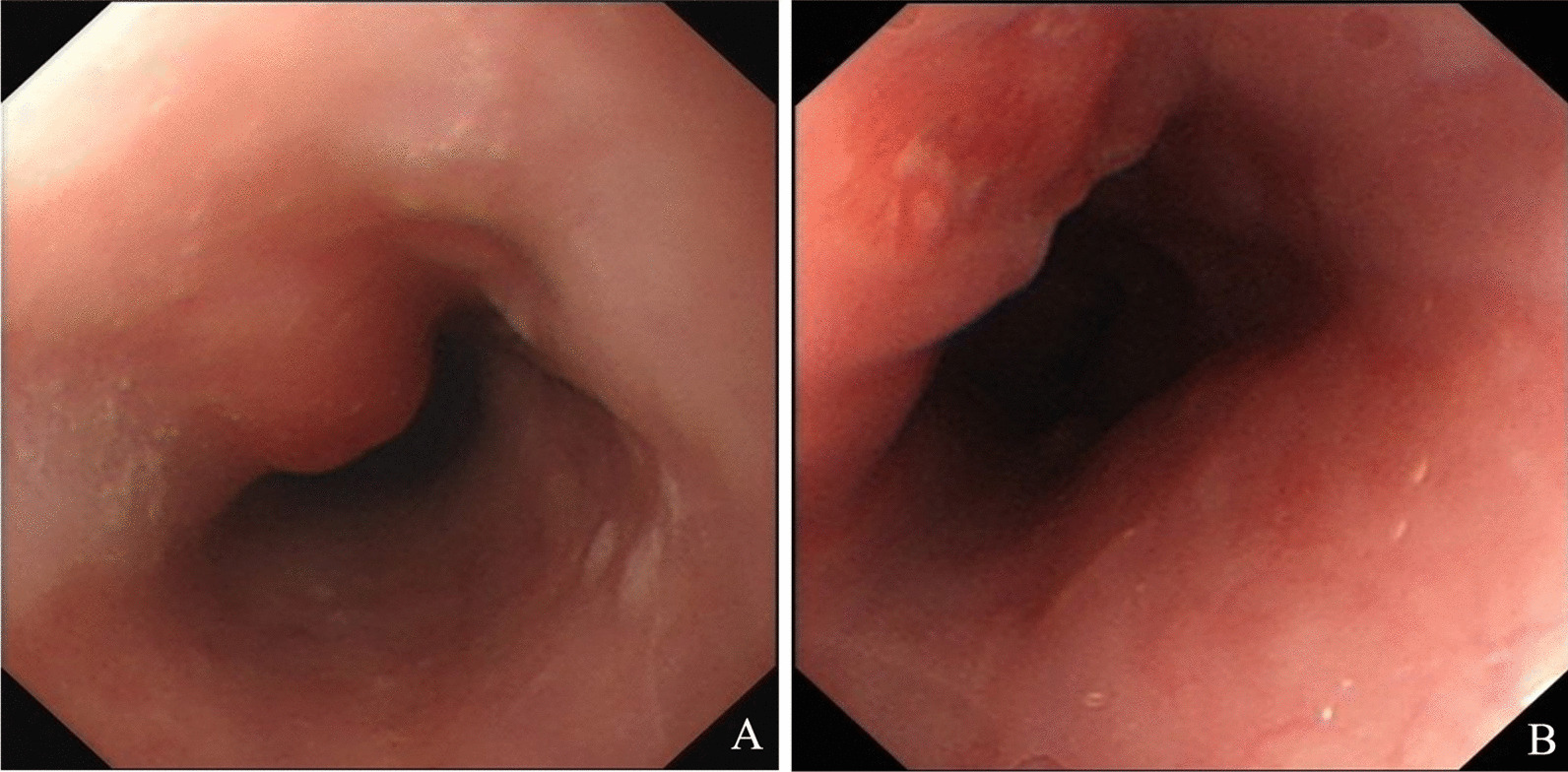
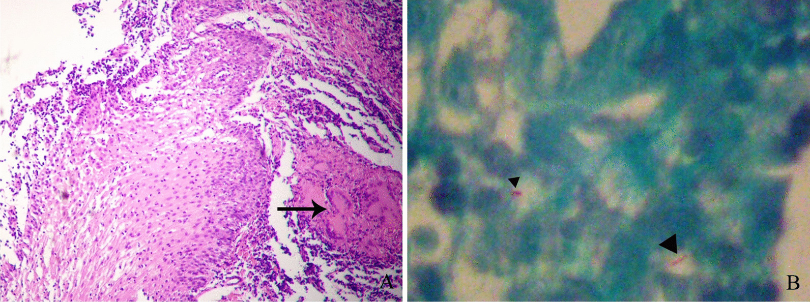
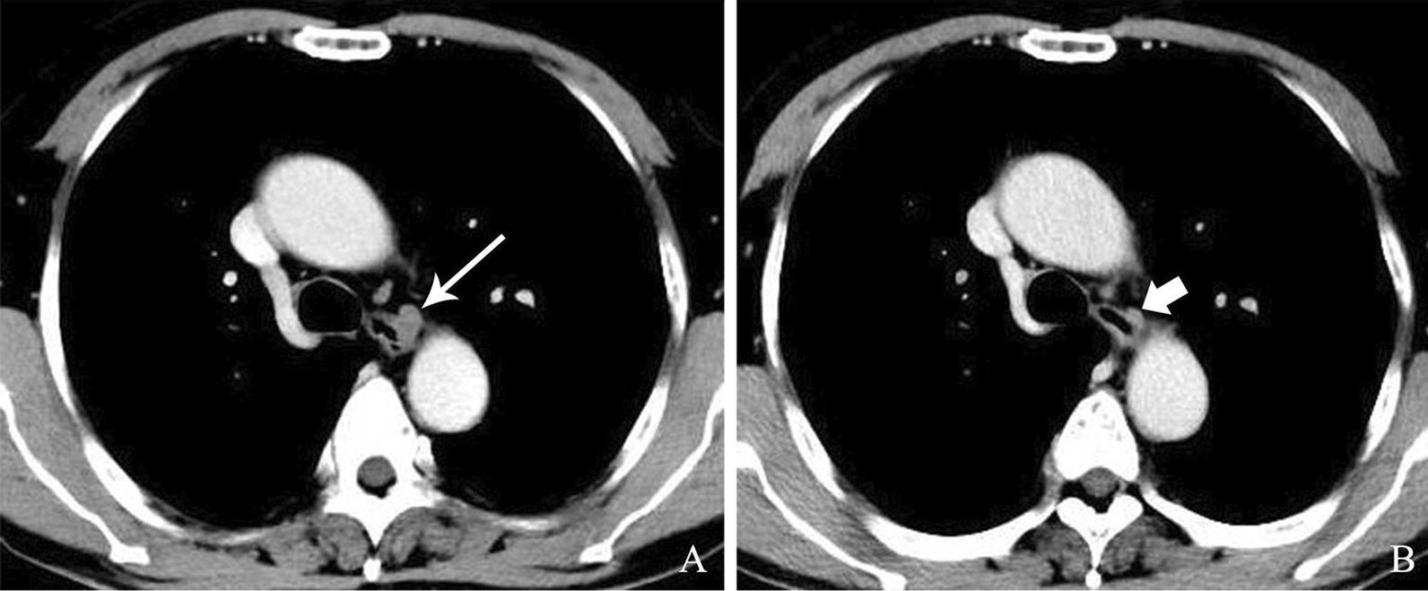
A 67-year-old male patient was admitted to the hospital due to "repeated chest pain for 1 month". Endoscopic ultrasonography and contrast-enhanced CT scans revealed a mass adjacent to the esophageal wall with unclear boundaries. Aspiration biopsy confirmed that esophageal tuberculosis was caused by nearby mediastinal tuberculous lymphadenitis. After a standard anti-tuberculosis treatment regimen, the patient achieved a favorable prognosis. The follow-up gastroesophageal endoscopy showed a sunken black lesion with elevated peripheral mucosa replacing the original tuberculous mass, which was thought to be anthracosis, a disease that rarely occurs in the esophagus.
The diagnosis of tuberculosis should be taken into consideration when a submucosal mass appears in the middle part of the esophagus. Endoscopic ultrasonography can effectively contribute to a definite diagnosis. Moreover, this is the first case of esophageal anthracosis observed only 1 year after the treatment of tuberculosis, indicating esophageal anthracosis can be a short-term disease. The traction of the reduction of tubercular mediastinal lymph nodes after anti-tuberculosis treatment may create a circumstance for pigmentation or dust deposition.
炭疽病通常被认为是由于在工作场所暴露于工业粉尘而导致的肺部疾病。食管炭疽病是一种相当罕见的现象,与结核病密切相关。此外,结核病累及食管也很少见。我们在此报告一例极其罕见的病例,在后续的胃肠内窥镜检查中发现一个肿块,中央有一个凹陷的黑色区域,周围黏膜呈凸起的环状,类似于恶性黑色素瘤。揭示患者的结核病病史最终避免了误诊或过度治疗。
一名 67 岁男性患者因“反复胸痛 1 个月”入院。内镜超声和增强 CT 扫描显示食管壁附近有一个边界不清的肿块。抽吸活检证实食管结核是由附近纵隔结核性淋巴结炎引起的。在接受标准抗结核治疗方案后,患者预后良好。后续的胃肠内窥镜检查显示,一个凹陷的黑色病变,周围黏膜隆起,取代了原来的结核肿块,这被认为是一种罕见发生在食管的炭疽病。
当食管中段出现黏膜下肿块时,应考虑结核病的诊断。内镜超声检查有助于明确诊断。此外,这是首例在结核病治疗仅 1 年后观察到的食管炭疽病,表明食管炭疽病可能是一种短期疾病。抗结核治疗后纵隔结核性淋巴结缩小的牵引可能为色素沉着或尘埃沉积创造了条件。