• 文献检索
  • 文档翻译
  • 深度研究
  • 学术资讯
  • Suppr Zotero 插件Zotero 插件
  • 邀请有礼
  • 套餐&价格
  • 历史记录
应用&插件
Suppr Zotero 插件Zotero 插件浏览器插件Mac 客户端Windows 客户端微信小程序
定价
高级版会员购买积分包购买API积分包
服务
文献检索文档翻译深度研究API 文档MCP 服务
关于我们
关于 Suppr公司介绍联系我们用户协议隐私条款
关注我们

Suppr 超能文献

核心技术专利:CN118964589B侵权必究
粤ICP备2023148730 号-1Suppr @ 2026

文献检索

告别复杂PubMed语法,用中文像聊天一样搜索,搜遍4000万医学文献。AI智能推荐,让科研检索更轻松。

立即免费搜索

文件翻译

保留排版,准确专业,支持PDF/Word/PPT等文件格式,支持 12+语言互译。

免费翻译文档

深度研究

AI帮你快速写综述,25分钟生成高质量综述,智能提取关键信息,辅助科研写作。

立即免费体验

尸检时发现的衰老性海马硬化在十多年前的磁共振成像(MRI)上就已很明显。

Hippocampal sclerosis of aging at post-mortem is evident on MRI more than a decade prior.

作者信息

Ortega-Cruz Diana, Eugenio Iglesias Juan, Rabano Alberto, Strange Bryan

机构信息

Laboratory for Clinical Neuroscience, Center for Biomedical Technology, Universidad Politécnica de Madrid, IdISSC, Madrid, Spain.

Alzheimer's Disease Research Unit, CIEN Foundation, Queen Sofia Foundation Alzheimer Center, Madrid, Spain.

出版信息

bioRxiv. 2023 Mar 10:2023.03.08.531683. doi: 10.1101/2023.03.08.531683.

DOI:10.1101/2023.03.08.531683
PMID:36945448
原文链接:https://pmc.ncbi.nlm.nih.gov/articles/PMC10028863/
Abstract

INTRODUCTION

Hippocampal sclerosis of aging (HS) is an important component of combined dementia neuropathology. However, the temporal evolution of its histologically-defined features is unknown. We investigated pre-mortem longitudinal hippocampal atrophy associated with HS, as well as with other dementia-associated pathologies.

METHODS

We analyzed hippocampal volumes from MRI segmentations in 64 dementia patients with longitudinal MRI follow-up and post-mortem neuropathological evaluation, including HS assessment in the hippocampal head and body.

RESULTS

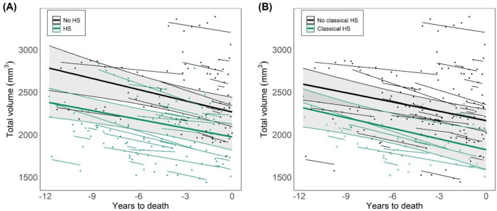
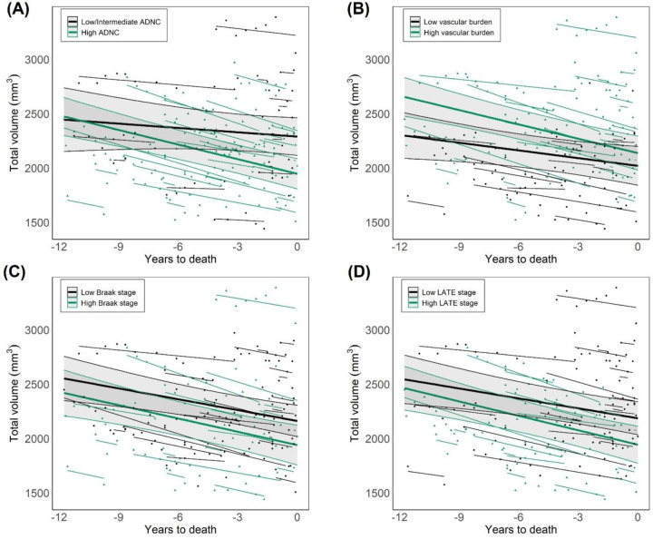
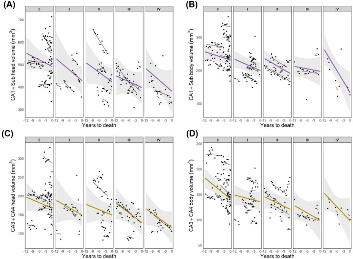
Significant HS-associated hippocampal volume changes were observed thoughout the evaluated timespan, up to 11.75 years before death. These changes were independent of age and Alzheimer’s Disease (AD) burden, and specifically driven by CA1 and subiculum. AD burden, but not HS, significantly associated with the rate of hippocampal atrophy.

DISCUSSION

HS-associated volume changes are detectable on MRI earlier than 10 years before death. These findings could contribute to the derivation of volumetric cut-offs for differentiation between HS and AD.

摘要

引言

衰老性海马硬化(HS)是混合性痴呆神经病理学的一个重要组成部分。然而,其组织学定义特征的时间演变尚不清楚。我们研究了与HS以及其他痴呆相关病理相关的生前纵向海马萎缩情况。

方法

我们分析了64例接受纵向MRI随访和死后神经病理学评估的痴呆患者MRI分割得到的海马体积,包括对海马头部和体部的HS评估。

结果

在长达死亡前11.75年的整个评估时间跨度内,观察到与HS相关的显著海马体积变化。这些变化与年龄和阿尔茨海默病(AD)负担无关,且具体由CA1和海马下托驱动。AD负担而非HS与海马萎缩率显著相关。

讨论

与HS相关的体积变化在死亡前10年以上即可通过MRI检测到。这些发现可能有助于得出区分HS和AD的体积临界值。

https://cdn.ncbi.nlm.nih.gov/pmc/blobs/bf46/10028863/517776dd06d7/nihpp-2023.03.08.531683v1-f0003.jpg
https://cdn.ncbi.nlm.nih.gov/pmc/blobs/bf46/10028863/524cd95d7023/nihpp-2023.03.08.531683v1-f0001.jpg
https://cdn.ncbi.nlm.nih.gov/pmc/blobs/bf46/10028863/18d2e550b55a/nihpp-2023.03.08.531683v1-f0002.jpg
https://cdn.ncbi.nlm.nih.gov/pmc/blobs/bf46/10028863/517776dd06d7/nihpp-2023.03.08.531683v1-f0003.jpg

相似文献

1
Hippocampal sclerosis of aging at post-mortem is evident on MRI more than a decade prior.尸检时发现的衰老性海马硬化在十多年前的磁共振成像(MRI)上就已很明显。
bioRxiv. 2023 Mar 10:2023.03.08.531683. doi: 10.1101/2023.03.08.531683.
2
Hippocampal sclerosis of aging at post-mortem is evident on MRI more than a decade prior.在尸检中,年龄相关性海马硬化症在 MRI 上的表现可追溯到 10 多年前。
Alzheimers Dement. 2023 Nov;19(11):5307-5315. doi: 10.1002/alz.13352. Epub 2023 Jun 27.
3
Utility of MRI in the identification of hippocampal sclerosis of aging.MRI 在老年海马硬化症识别中的应用。
Alzheimers Dement. 2021 May;17(5):847-855. doi: 10.1002/alz.12241. Epub 2020 Dec 7.
4
Longitudinal hippocampal atrophy in hippocampal sclerosis of aging.衰老性海马硬化中的海马纵向萎缩
Aging Brain. 2023 Aug 12;4:100092. doi: 10.1016/j.nbas.2023.100092. eCollection 2023.
5
Manual Hippocampal Subfield Segmentation Using High-Field MRI: Impact of Different Subfields in Hippocampal Volume Loss of Temporal Lobe Epilepsy Patients.使用高场磁共振成像进行海马亚区手动分割:不同亚区对颞叶癫痫患者海马体积损失的影响
Front Neurol. 2018 Nov 20;9:927. doi: 10.3389/fneur.2018.00927. eCollection 2018.
6
A novel histological staging of hippocampal sclerosis that is evident in gray matter loss in vivo.一种新的海马硬化组织学分期,在体内表现为灰质丢失。
Alzheimers Dement. 2023 Jul;19(7):3028-3040. doi: 10.1002/alz.12942. Epub 2023 Jan 24.
7
Correlating hippocampal and amygdala volumes with neuropathological burden in neurodegenerative diseases using 7T postmortem MRI.使用7T尸检MRI将海马体和杏仁核体积与神经退行性疾病中的神经病理学负担相关联。
medRxiv. 2024 May 15:2024.05.15.24307354. doi: 10.1101/2024.05.15.24307354.
8
Characterization of hippocampal sclerosis of aging and its association with other neuropathologic changes and cognitive deficits in the oldest-old.老年海马硬化的特征及其与最老年人群中其他神经病理学变化和认知缺陷的关系。
Acta Neuropathol. 2023 Sep;146(3):415-432. doi: 10.1007/s00401-023-02606-9. Epub 2023 Jun 29.
9
MRI shows more severe hippocampal atrophy and shape deformation in hippocampal sclerosis than in Alzheimer's disease.磁共振成像显示,与阿尔茨海默病相比,海马硬化中的海马萎缩和形态变形更为严重。
Int J Alzheimers Dis. 2011 Apr 11;2011:483972. doi: 10.4061/2011/483972.
10
Hippocampal sclerosis of aging is a key Alzheimer's disease mimic: clinical-pathologic correlations and comparisons with both alzheimer's disease and non-tauopathic frontotemporal lobar degeneration.老年海马硬化是阿尔茨海默病的重要模拟物:临床病理相关性及与阿尔茨海默病和非tau 蛋白病性额颞叶变性的比较。
J Alzheimers Dis. 2014;39(3):691-702. doi: 10.3233/JAD-131880.

本文引用的文献

1
SynthSeg: Segmentation of brain MRI scans of any contrast and resolution without retraining.SynthSeg:无需重新训练即可对任何对比度和分辨率的脑 MRI 扫描进行分割。
Med Image Anal. 2023 May;86:102789. doi: 10.1016/j.media.2023.102789. Epub 2023 Feb 25.
2
Robust machine learning segmentation for large-scale analysis of heterogeneous clinical brain MRI datasets.用于大规模分析异质临床脑 MRI 数据集的稳健机器学习分割。
Proc Natl Acad Sci U S A. 2023 Feb 28;120(9):e2216399120. doi: 10.1073/pnas.2216399120. Epub 2023 Feb 21.
3
A novel histological staging of hippocampal sclerosis that is evident in gray matter loss in vivo.
一种新的海马硬化组织学分期,在体内表现为灰质丢失。
Alzheimers Dement. 2023 Jul;19(7):3028-3040. doi: 10.1002/alz.12942. Epub 2023 Jan 24.
4
Neuropathological correlates of neuropsychiatric symptoms in dementia.痴呆患者神经精神症状的神经病理学相关性。
Alzheimers Dement. 2023 Apr;19(4):1372-1382. doi: 10.1002/alz.12765. Epub 2022 Sep 23.
5
Cerebrospinal Fluid Biomarkers in Autopsy-Confirmed Alzheimer Disease and Frontotemporal Lobar Degeneration.尸检证实的阿尔茨海默病和额颞叶变性中的脑脊液生物标志物。
Neurology. 2022 Mar 15;98(11):e1137-e1150. doi: 10.1212/WNL.0000000000200040. Epub 2022 Feb 16.
6
The vascular risk factors and vascular neuropathology in subjects with autopsy-confirmed dementia with Lewy bodies.尸检确诊的路易体痴呆患者的血管危险因素和血管神经病理学。
Int J Geriatr Psychiatry. 2022 Mar;37(3). doi: 10.1002/gps.5683.
7
Distinct characteristics of limbic-predominant age-related TDP-43 encephalopathy in Lewy body disease.路易体病中以边缘系统为主的与年龄相关的 TDP-43 脑病的特征。
Acta Neuropathol. 2022 Jan;143(1):15-31. doi: 10.1007/s00401-021-02383-3. Epub 2021 Dec 2.
8
Hippocampal Sclerosis in Frontotemporal Dementia: When Vascular Pathology Meets Neurodegeneration.额颞叶痴呆中的海马硬化:血管病变与神经退行性变相遇时。
J Neuropathol Exp Neurol. 2021 Mar 22;80(4):313-324. doi: 10.1093/jnen/nlab010.
9
Utility of MRI in the identification of hippocampal sclerosis of aging.MRI 在老年海马硬化症识别中的应用。
Alzheimers Dement. 2021 May;17(5):847-855. doi: 10.1002/alz.12241. Epub 2020 Dec 7.
10
Hippocampal sclerosis, TDP-43, and the duration of the symptoms of dementia of AD patients.海马硬化症、TDP-43 与 AD 患者痴呆症状持续时间的关系。
Ann Clin Transl Neurol. 2020 Sep;7(9):1546-1556. doi: 10.1002/acn3.51135. Epub 2020 Jul 31.