Department of Radiology, Pamukkale University Faculty of Medicine, Denizli, Turkey.
Department of Radiology, Hacettepe University Faculty of Medicine, Ankara, Turkey.
Diagn Interv Radiol. 2023 Jan 31;29(1):68-79. doi: 10.4274/dir.2022.221419. Epub 2023 Jan 2.
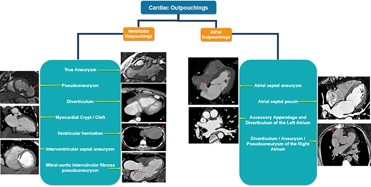
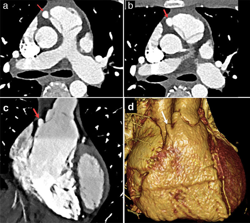
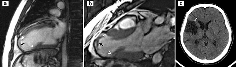
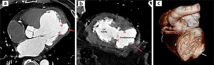
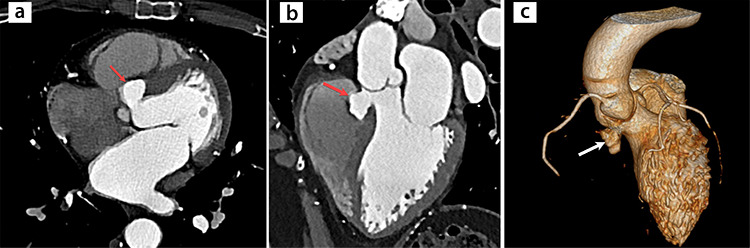
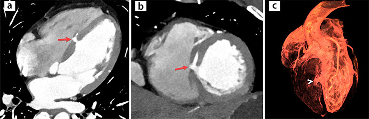
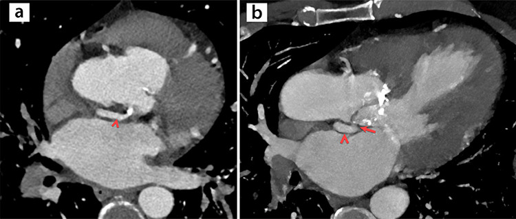
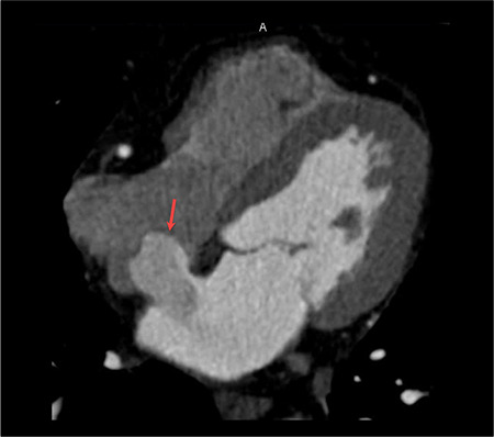
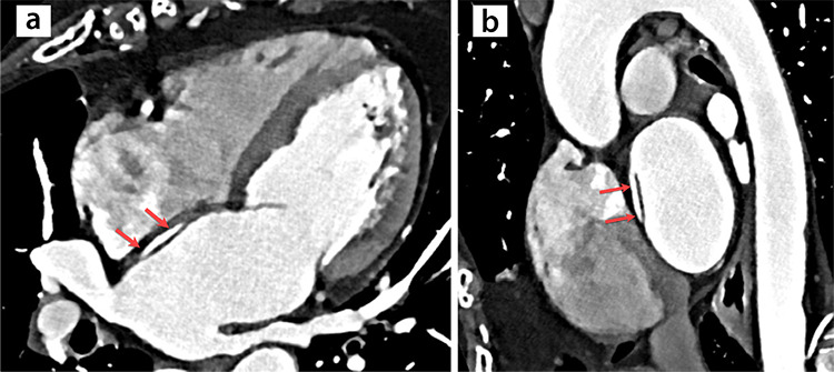
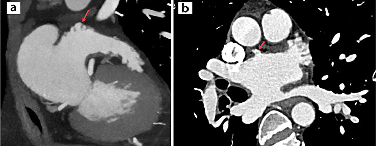
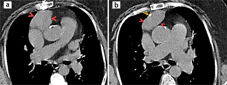
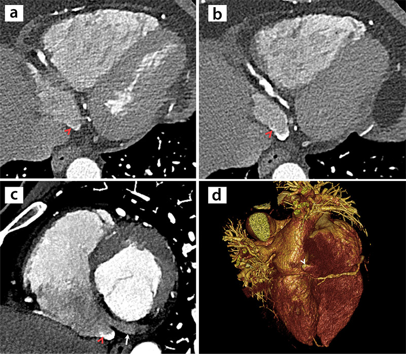
A cardiac outpouching (CO) is a protrusion in a heart chamber's internal anatomical lining. Most COs are clinically insignificant, but some are of vital importance, requiring immediate surgery. Cross-sectional imaging findings of COs, such as location, morphology, size, and accompanying wall motion abnormalities, play an essential role in determining the correct diagnosis and appropriate clinical management. Therefore, radiologists should be familiar with them. This article reviews the key cross-sectional imaging findings and differential diagnoses of COs.
心腔膨出(CO)是心脏腔室内膜的一种突起。大多数 CO 无临床意义,但有些 CO 至关重要,需要立即手术。CO 的横断面成像表现,如位置、形态、大小和伴随的壁运动异常,对于确定正确的诊断和适当的临床管理至关重要。因此,放射科医生应该熟悉这些表现。本文复习了 CO 的关键横断面成像表现和鉴别诊断。