Department of Neurosurgery, The Second Affiliated Hospital of Nanchang University, Jiangxi, 330006, Nanchang, P. R. China.
Jiangxi Key Laboratory of Neurological Tumors and Cerebrovascular Diseases, Jiangxi, 330006, Nanchang, P. R. China.
J Exp Clin Cancer Res. 2023 Mar 30;42(1):77. doi: 10.1186/s13046-023-02640-1.
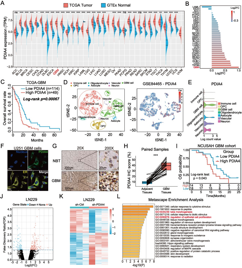
Increasing evidence has revealed the key activity of protein disulfide isomerase A4 (PDIA4) in the endoplasmic reticulum stress (ERS) response. However, the role of PDIA4 in regulating glioblastoma (GBM)-specific pro-angiogenesis is still unknown.
The expression and prognostic role of PDIA4 were analyzed using a bioinformatics approach and were validated in 32 clinical samples and follow-up data. RNA-sequencing was used to search for PDIA4-associated biological processes in GBM cells, and proteomic mass spectrum (MS) analysis was used to screen for potential PDIA4 substrates. Western blotting, real-time quantitative polymerase chain reaction (RT-qPCR), and enzyme-linked immunosorbent assays (ELISA) were used to measure the levels of the involved factors. Cell migration and tube formation assays determined the pro-angiogenesis activity of PDIA4 in vitro. An intracranial U87 xenograft GBM animal model was constructed to evaluate the pro-angiogenesis role of PDIA4 in vivo.
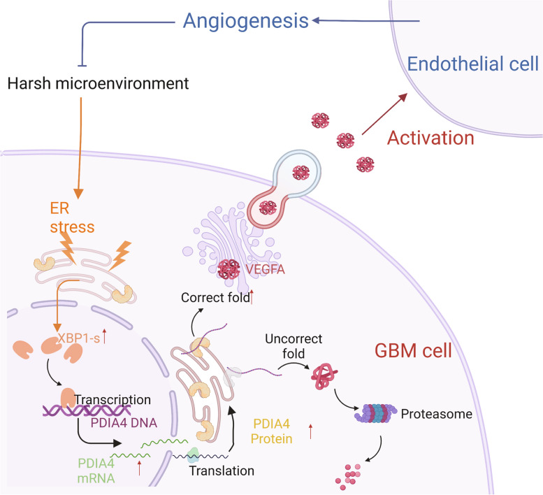
Aberrant overexpression of PDIA4 was associated with a poor prognosis in patients with GBM, although PDIA4 could also functionally regulate intrinsic GBM secretion of vascular endothelial growth factor-A (VEGF-A) through its active domains of Cys-X-X-Cys (CXXC) oxidoreductase. Functionally, PDIA4 exhibits pro-angiogenesis activity both in vitro and in vivo, and can be upregulated by ERS through transcriptional regulation of X-box binding protein 1 (XBP1). The XBP1/PDIA4/VEGFA axis partially supports the mechanism underlying GBM cell survival under ER stress. Further, GBM cells with higher expression of PDIA4 showed resistance to antiangiogenic therapy in vivo.
Our findings revealed the pro-angiogenesis role of PDIA4 in GBM progression and its potential impact on GBM survival under a harsh microenvironment. Targeting PDIA4 might help to improve the efficacy of antiangiogenic therapy in patients with GBM.
越来越多的证据表明,蛋白质二硫键异构酶 A4(PDIA4)在内质网应激(ERS)反应中的关键活性。然而,PDIA4 在调节胶质母细胞瘤(GBM)特异性促血管生成中的作用尚不清楚。
使用生物信息学方法分析 PDIA4 的表达和预后作用,并在 32 个临床样本和随访数据中进行验证。RNA 测序用于搜索 GBM 细胞中与 PDIA4 相关的生物学过程,蛋白质组学质谱(MS)分析用于筛选潜在的 PDIA4 底物。Western blot、实时定量聚合酶链反应(RT-qPCR)和酶联免疫吸附试验(ELISA)用于测量相关因素的水平。细胞迁移和管形成试验测定了 PDIA4 在体外的促血管生成活性。构建颅内 U87 异种移植 GBM 动物模型,体内评价 PDIA4 的促血管生成作用。
PDIA4 的异常过表达与 GBM 患者的不良预后相关,尽管 PDIA4 还可以通过其 Cys-X-X-Cys(CXXC)氧化还原酶的活性域功能调节内在 GBM 血管内皮生长因子-A(VEGF-A)的分泌。在功能上,PDIA4 在体外和体内均具有促血管生成活性,并且可以通过 X 盒结合蛋白 1(XBP1)的转录调节而上调。XBP1/PDIA4/VEGFA 轴部分支持了 ERS 下 GBM 细胞存活的机制。此外,表达 PDIA4 较高的 GBM 细胞在体内对抗血管生成治疗表现出耐药性。
我们的研究结果揭示了 PDIA4 在 GBM 进展中的促血管生成作用及其在恶劣微环境下对 GBM 存活的潜在影响。靶向 PDIA4 可能有助于提高 GBM 患者抗血管生成治疗的疗效。