Turku PET Centre, University of Turku, Turku University Hospital, Kiinamyllynkatu 4-8, 20520, Turku, Finland.
Department of Psychiatry and Neurochemistry, Institute of Neuroscience & Physiology, the Sahlgrenska Academy at the University of Gothenburg, Mölndal, Sweden.
Alzheimers Res Ther. 2023 Apr 4;15(1):71. doi: 10.1186/s13195-023-01209-6.
Neuroinflammation, characterized by increased reactivity of microglia and astrocytes in the brain, is known to be present at various stages of the Alzheimer's disease (AD) continuum. However, its presence and relationship with amyloid pathology in cognitively normal at-risk individuals is less clear. Here, we used positron emission tomography (PET) and blood biomarker measurements to examine differences in neuroinflammation and beta-amyloid (Aβ) and their association in cognitively unimpaired homozygotes, heterozygotes, or non-carriers of the APOE ε4 allele, the strongest genetic risk for sporadic AD.
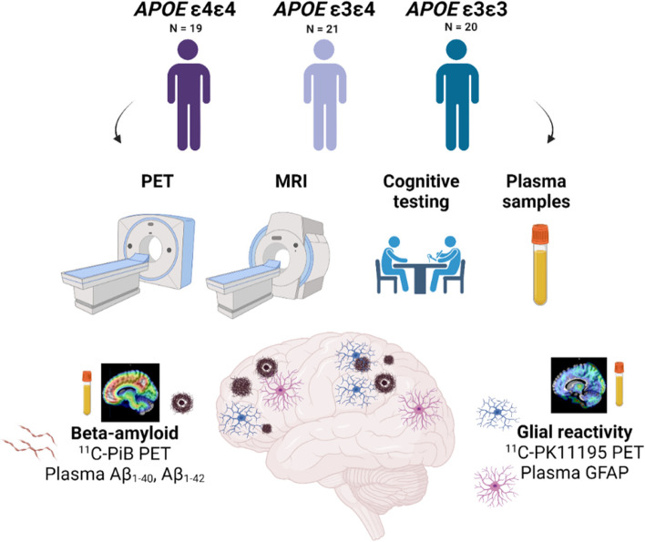
Sixty 60-75-year-old APOE ε4 homozygotes (n = 19), heterozygotes (n = 21), and non-carriers (n = 20) were recruited in collaboration with the local Auria biobank. The participants underwent C-PK11195 PET (targeting 18-kDa translocator protein, TSPO), C-PiB PET (targeting Aβ), brain MRI, and neuropsychological testing including a preclinical cognitive composite (APCC). C-PK11195 distribution volume ratios and C-PiB standardized uptake value ratios (SUVRs) were calculated for regions typical for early Aβ accumulation in AD. Blood samples were drawn for measuring plasma glial fibrillary acidic protein (GFAP) and plasma Aβ.
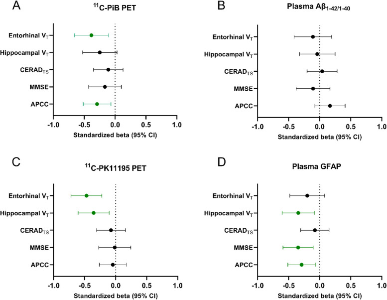
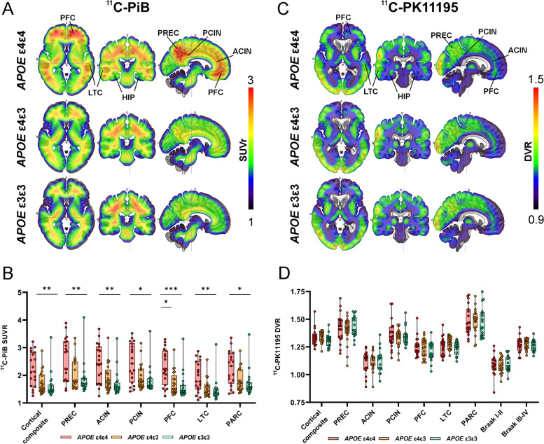
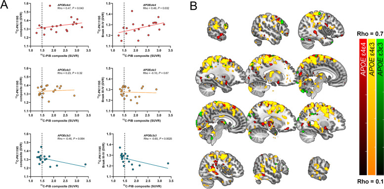
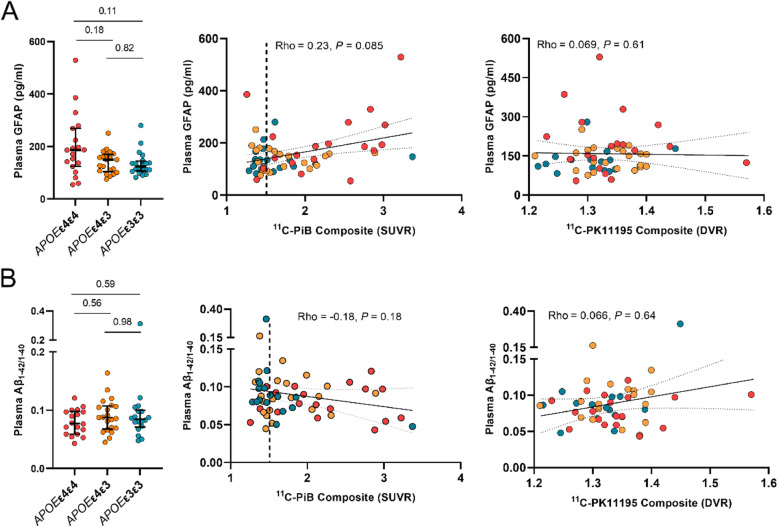
In our cognitively unimpaired sample, cortical C-PiB-binding increased according to APOE ε4 gene dose (median composite SUVR 1.47 (range 1.38-1.66) in non-carriers, 1.55 (1.43-2.02) in heterozygotes, and 2.13 (1.61-2.83) in homozygotes, P = 0.002). In contrast, cortical composite C-PK11195-binding did not differ between the APOE ε4 gene doses (P = 0.27) or between Aβ-positive and Aβ-negative individuals (P = 0.81) and associated with higher Aβ burden only in APOE ε4 homozygotes (Rho = 0.47, P = 0.043). Plasma GFAP concentration correlated with cortical C-PiB (Rho = 0.35, P = 0.040), but not C-PK11195-binding (Rho = 0.13, P = 0.47) in Aβ-positive individuals. In the total cognitively unimpaired population, both higher composite C-PK11195-binding and plasma GFAP were associated with lower hippocampal volume, whereas elevated C-PiB-binding was associated with lower APCC scores.
Only Aβ burden measured by PET, but not markers of neuroinflammation, differed among cognitively unimpaired elderly with different APOE ε4 gene dose. However, APOE ε4 gene dose seemed to modulate the association between neuroinflammation and Aβ.
神经炎症的特征是大脑中小胶质细胞和星形胶质细胞的反应性增加,已知在阿尔茨海默病(AD)连续体的各个阶段都存在。然而,在认知正常的高危人群中,其存在及其与淀粉样蛋白病理学的关系尚不清楚。在这里,我们使用正电子发射断层扫描(PET)和血液生物标志物测量来检查认知正常的纯合子、杂合子或非载脂蛋白 E4 等位基因(APOE ε4)个体中神经炎症和β-淀粉样蛋白(Aβ)的差异及其相关性,APOE ε4 是散发性 AD 的最强遗传风险因素。
60 名 60-75 岁的 APOE ε4 纯合子(n=19)、杂合子(n=21)和非携带者(n=20)在当地奥里亚生物库的合作下招募。参与者接受 C-PK11195 PET(靶向 18kDa 转位蛋白,TSPO)、C-PiB PET(靶向 Aβ)、脑 MRI 和神经心理学测试,包括临床前认知复合(APCC)。计算了 Aβ 早期积累的 AD 典型区域的 C-PK11195 分布容积比和 C-PiB 标准化摄取值比(SUVR)。抽取血液样本测量血浆神经胶质纤维酸性蛋白(GFAP)和血浆 Aβ。
在我们认知正常的样本中,根据 APOE ε4 基因剂量,皮质 C-PiB 结合增加(非携带者复合 SUVR 中位数为 1.47(范围 1.38-1.66),杂合子为 1.55(1.43-2.02),纯合子为 2.13(1.61-2.83),P=0.002)。相比之下,APOE ε4 基因剂量之间的皮质复合 C-PK11195 结合没有差异(P=0.27),Aβ 阳性和 Aβ 阴性个体之间也没有差异(P=0.81),仅在 APOE ε4 纯合子中与更高的 Aβ 负担相关(Rho=0.47,P=0.043)。在 Aβ 阳性个体中,血浆 GFAP 浓度与皮质 C-PiB 相关(Rho=0.35,P=0.040),但与 C-PK11195 结合无关(Rho=0.13,P=0.47)。在整个认知正常人群中,较高的复合 C-PK11195 结合和血浆 GFAP 均与海马体积降低相关,而较高的 C-PiB 结合与 APCC 评分降低相关。
在不同 APOE ε4 基因剂量的认知正常老年人中,只有通过 PET 测量的 Aβ 负担而不是神经炎症标志物存在差异。然而,APOE ε4 基因剂量似乎调节了神经炎症与 Aβ 之间的关联。