Department of Radiology, Faculty of Medicine, Kagawa University, 1750-1 Ikenobe, Miki-Cho, Kita-Gun, Kagawa, 761-0793, Japan.
Department of Radiology, Diagnostic Imaging Center, Utazu Hospital, Utazu-Cho, Ayauta-Gun, Kagawa, Japan.
Jpn J Radiol. 2023 Sep;41(9):965-972. doi: 10.1007/s11604-023-01424-z. Epub 2023 Apr 11.
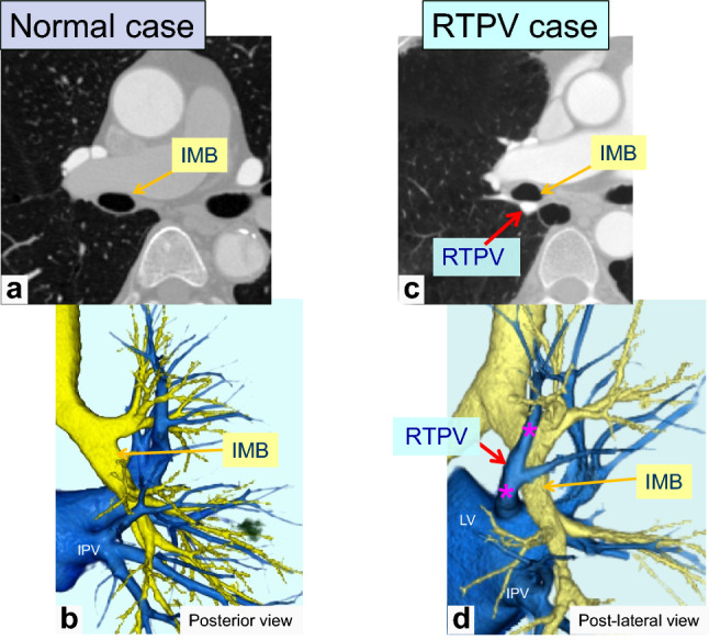
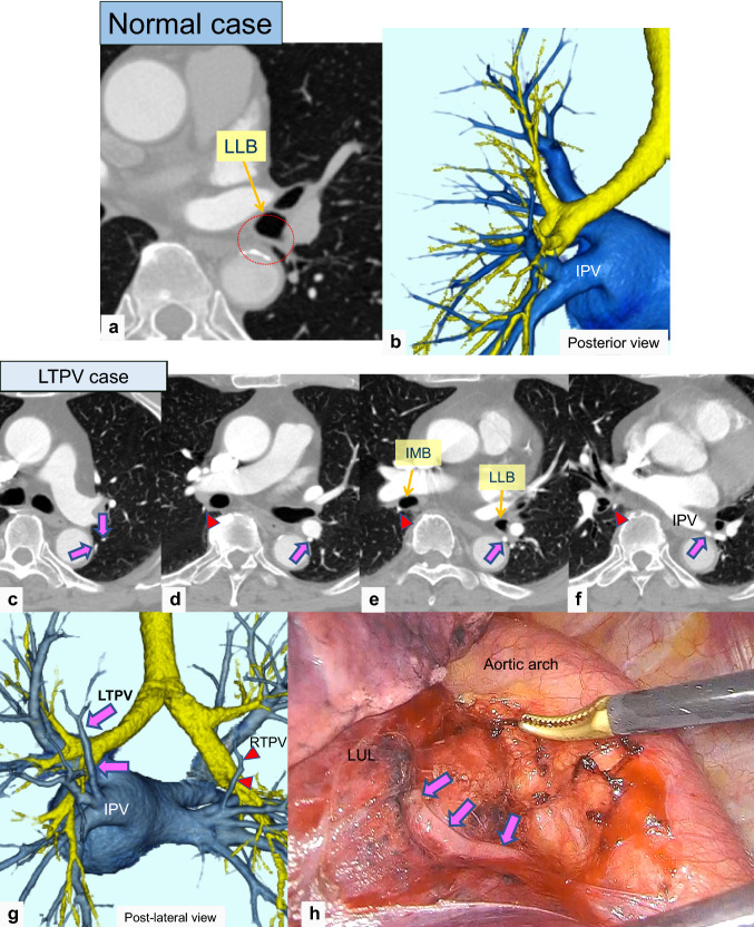
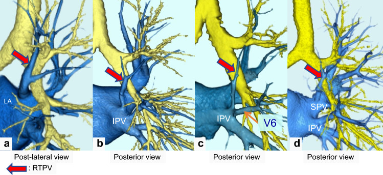
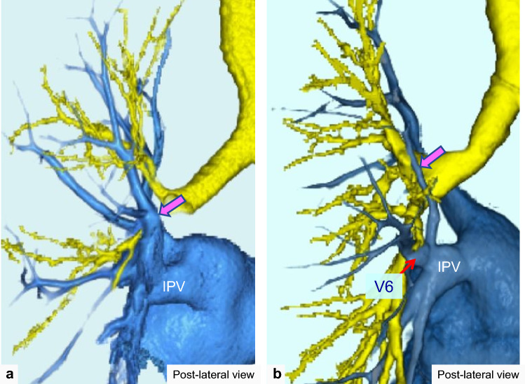
The right top pulmonary vein (RTPV) is defined as an anomalous branch of the right superior PV (SPV) draining into the PV or left atrium (LA). Several previous reports have described the RTPV, but only a few have mentioned the left top PV (LTPV). The present study aimed to evaluate the branching patterns of the RTPV and LTPV using thin-section CT images and three-dimensional CT angiography (3D-CTA).
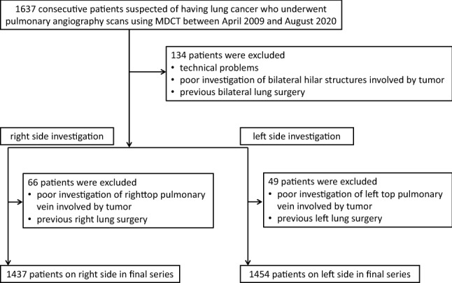
This study included 1437 consecutive patients for evaluation of the right side and 1454 consecutive patients for the left side who were suspected of lung cancer and underwent CTA. We assessed the presence of each RTPV and LTPV and their branching patterns on the CTA images. When the RTPV or LTPV was identified, the maximum short-axis diameter was measured.
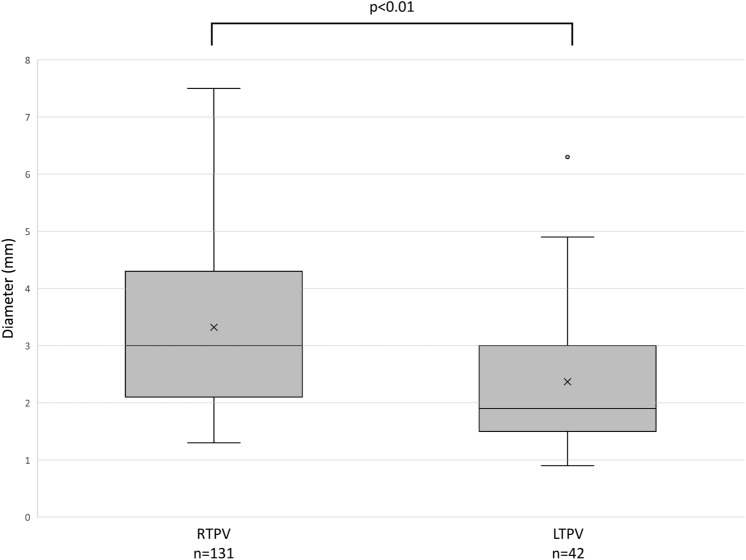
RTPV was found in 9.1% (131/1437), whereas LTPV was found in 2.9% (42/1454) of the patients. RTPV was also observed in 17.1% (7/41) of LTPV cases, except for one case in which the right side could not be evaluated. The most common RTPV inflow site was the right inferior PV (IPV) in 64.9% (85/131) of the patients, whereas that of the LTPV was the left IPV in 100.0% (42/42) of the patients. The mean diameter of the RTPV and LTPV was 3.3 mm (range, 1.3-7.5 mm) and 2.4 mm (range, 0.9-6.3 mm), respectively (P < 0.01).
The top PV branching pattern variations can be evaluated using thin-section CT and 3D-CTA images. RTPV is not a rare finding, and LTPV should also be identified in lung cancer cases scheduled for resection.
右上肺静脉(RTPV)定义为右优势型肺静脉(RSPV)的异常分支,引流至肺静脉或左心房(LA)。之前有几项研究描述了 RTPV,但只有少数研究提到了左上肺静脉(LTPV)。本研究旨在使用薄层 CT 图像和三维 CT 血管造影(3D-CTA)评估 RTPV 和 LTPV 的分支模式。
本研究纳入了 1437 例连续疑似肺癌患者的右侧和 1454 例连续疑似肺癌患者的左侧 CTA 评估,共 2891 例。我们在 CTA 图像上评估了每支 RTPV 和 LTPV 的存在及其分支模式。当识别出 RTPV 或 LTPV 时,测量其最大短轴直径。
9.1%(131/1437)的患者存在 RTPV,2.9%(42/1454)的患者存在 LTPV。在 17.1%(7/41)的 LTPV 病例中也观察到 RTPV,除了 1 例右侧无法评估。64.9%(85/131)的 RTPV 患者的 RTPV 流入部位最常见于右下肺静脉(IPV),而 100.0%(42/42)的 LTPV 患者的流入部位为左上肺静脉(IPV)。RTPV 和 LTPV 的平均直径分别为 3.3mm(范围 1.3-7.5mm)和 2.4mm(范围 0.9-6.3mm)(P<0.01)。
使用薄层 CT 和 3D-CTA 图像可以评估顶级 PV 分支模式的变化。RTPV 并不罕见,在计划进行肺切除术的肺癌患者中也应识别 LTPV。