Department of Thoracic, and Endocrine Surgery and Oncology, Institute of Biomedical Sciences, The University of Tokushima Graduate School, 3-18-15, Kuramoto-cho, Tokushima, 770-8503, Japan.
J Cardiothorac Surg. 2021 Jan 21;16(1):15. doi: 10.1186/s13019-021-01392-3.
A displaced left B accompanied by an anomalous pulmonary vein is a rare condition involving complex structures. There is a risk of unexpected injuries to bronchi and blood vessels when patients with such anomalies undergo surgery for lung cancer.
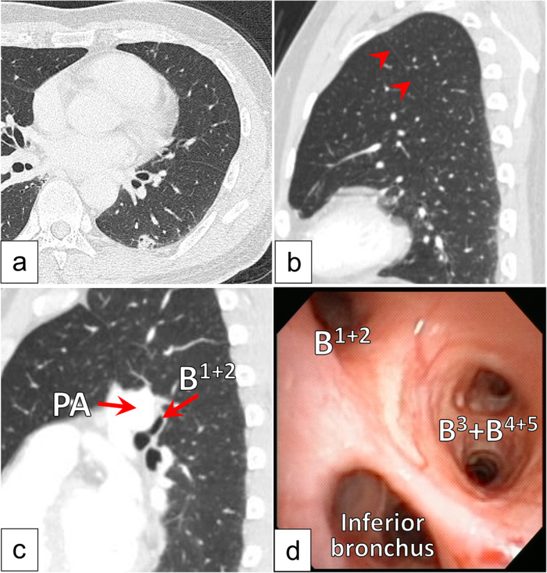
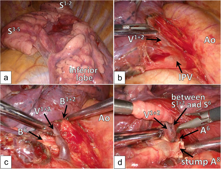
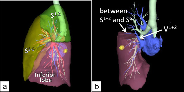
A 59-year-old male with suspected lung cancer in the left lower lobe was scheduled to undergo surgery. Chest computed tomography revealed a displaced B and hyperlobulation between S and S, while the interlobar fissure between S and S was completely fused. Three-dimensional computed tomography (3D-CT) revealed an anomalous V joining the left inferior pulmonary vein and a branch of the V running between S and S. We performed left lower lobectomy via video-assisted thoracic surgery, while taking care with the abovementioned anatomical structures. The strategy employed in this operation was to preserve V and confirm the locations of B and B when dividing the fissure.
The aim of the surgical procedure performed in this case was to divide the fissure between S and the inferior lobe to reduce the risk of an unexpected bronchial injury. 3D-CT helps surgeons to understand the stereoscopic positional relationships among anatomical structures.
伴有异常肺静脉的左 B 移位是一种涉及复杂结构的罕见情况。当患有此类异常的患者因肺癌接受手术时,存在意外损伤支气管和血管的风险。
一名 59 岁男性,因左下肺疑似肺癌而接受手术。胸部计算机断层扫描显示 S 和 S 之间的 B 移位和过度膨胀,而 S 和 S 之间的叶间裂完全融合。三维计算机断层扫描(3D-CT)显示 V 异常连接左下肺静脉和 V 的一支在 S 和 S 之间运行。我们通过电视辅助胸腔手术进行了左下肺叶切除术,同时注意了上述解剖结构。该手术采用的策略是保留 V,并在分隔裂时确认 B 和 B 的位置。
本例手术的目的是分隔 S 和下叶之间的裂,以降低意外支气管损伤的风险。3D-CT 有助于外科医生了解解剖结构之间的立体位置关系。