Yi Bingcheng, Zhou Boya, Song Zhenfeng, Yu Lei, Wang Wenbo, Liu Wei
Department of Plastic and Reconstructive Surgery, Shanghai Key Laboratory of Tissue Engineering, Shanghai Ninth People's Hospital, Shanghai Jiao Tong University School of Medicine, Shanghai, 200011, China.
Department of Vascular Surgery, Shanghai Ninth People's Hospital, Shanghai Jiao Tong University School of Medicine, Shanghai, 200011, China.
Bioact Mater. 2022 Jul 14;25:657-676. doi: 10.1016/j.bioactmat.2022.07.010. eCollection 2023 Jul.
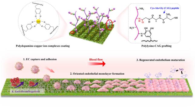
Native-like endothelium regeneration is a prerequisite for material-guided small-diameter vascular regeneration. In this study, a novel strategy is proposed to achieve phase-adjusted endothelial healing by step-wise modification of parallel-microgroove-patterned (i.e., micropatterned) nanofibers with polydopamine-copper ion (PDA-Cu) complexes, polylysine (PLys) molecules, and Cys-Ala-Gly (CAG) peptides (CAG@PLys@PDA-Cu). Using electrospun poly(-lactide--caprolactone) random nanofibers as the demonstrating biomaterial, step-wise modification of CAG@PLys@PDA-Cu significantly enhanced substrate wettability and protein adsorption, exhibited an excellent antithrombotic surface and outstanding phase-adjusted capacity of endothelium regeneration involving cell adhesion, endothelial monolayer formation, and the regenerated endothelium maturation. Upon implantation for segmental replacement of rabbit carotid arteries, CAG@PLys@PDA-Cu modified grafts (2 mm inner diameter) with micropatterns on inner surface effectively accelerated native-like endothelium regeneration within 1 week, with less platelet aggregates and inflammatory response compared to those on non-modified grafts. Prolonged observations at 6- and 12-weeks post-implantation demonstrated a positive vascular remodeling with almost fully covered endothelium and mature smooth muscle layer in the modified vascular grafts, accompanied with well-organized extracellular matrix. By contrast, non-modified vascular grafts induced a disorganized tissue formation with a high risk of thrombogenesis. In summary, step-wise modification of CAG@PLys@PDA-Cu on micropatterned nanofibers can significantly promote endothelial healing without inflicting thrombosis, thus confirming a novel strategy for developing functional vascular grafts or other blood-contacting materials/devices.
类天然内皮再生是材料引导下小口径血管再生的前提条件。在本研究中,我们提出了一种新策略,通过用聚多巴胺 - 铜离子(PDA-Cu)复合物、聚赖氨酸(PLys)分子和半胱氨酸 - 丙氨酸 - 甘氨酸(CAG)肽(CAG@PLys@PDA-Cu)对平行微槽图案化(即微图案化)纳米纤维进行逐步修饰,实现相位调整的内皮愈合。以静电纺丝聚(- 丙交酯 - 己内酯)无规纳米纤维作为示范生物材料,CAG@PLys@PDA-Cu的逐步修饰显著提高了底物润湿性和蛋白质吸附能力,展现出优异的抗血栓表面以及在涉及细胞黏附、内皮单层形成和再生内皮成熟方面出色的相位调整内皮再生能力。在植入兔颈动脉进行节段置换后,内表面带有微图案的CAG@PLys@PDA-Cu修饰移植物(内径2毫米)在1周内有效加速了类天然内皮再生,与未修饰移植物相比,血小板聚集和炎症反应更少。植入后6周和12周的长期观察表明,修饰后的血管移植物呈现出积极的血管重塑,内皮几乎完全覆盖,平滑肌层成熟,同时伴有组织良好的细胞外基质。相比之下,未修饰的血管移植物诱导形成无序的组织,血栓形成风险高。总之,在微图案化纳米纤维上对CAG@PLys@PDA-Cu进行逐步修饰可显著促进内皮愈合而不引发血栓形成,从而证实了一种开发功能性血管移植物或其他血液接触材料/装置的新策略。