Mahawar Rajat, Dharamshi Jay D, Shinde Raju K, Rathi Chetna
General Surgery, Jawaharlal Nehru Medical College, Datta Meghe Institute of Higher Education and Research, Wardha, IND.
Urosurgery, Jawaharlal Nehru Medical College, Datta Meghe Institute of Higher Education and Research, Wardha, IND.
Cureus. 2023 Mar 14;15(3):e36141. doi: 10.7759/cureus.36141. eCollection 2023 Mar.
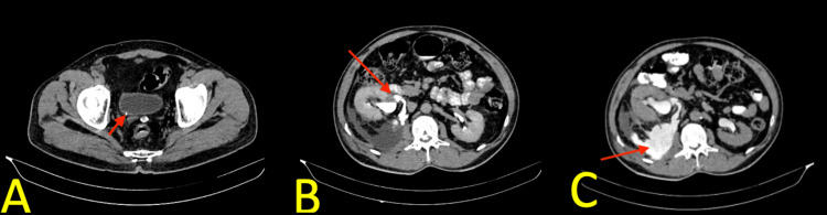
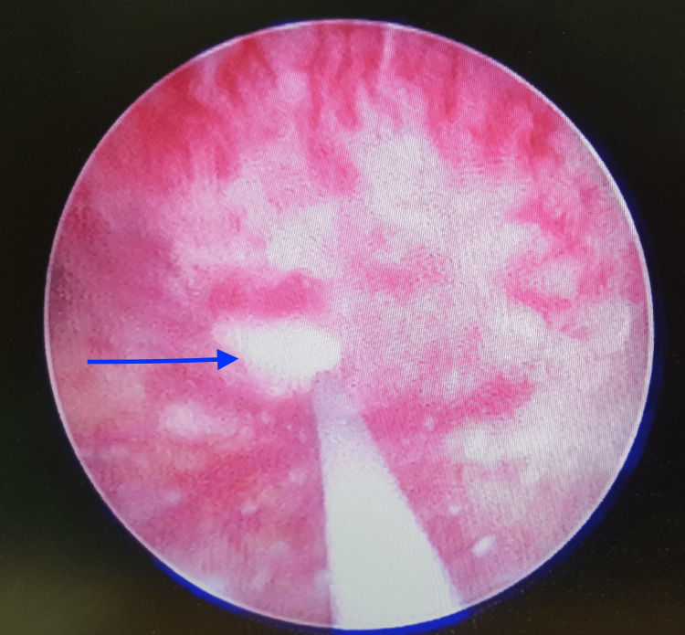
Spontaneous rupture of the renal pelvis (SRRP) with urine extravasation is rare. This condition is primarily associated with an obstructing ureteric calculus. It creates a diagnostic dilemma, especially when the clinical diagnosis can be inconsistent. Herein, we report a 49-year-old male patient who presented with abdominal pain for the past three days and was diagnosed with acute appendicitis. A computed tomography (CT) scan revealed a right renal pelvis rupture and urinoma secondary to an obstructive 4 mm ureterovesical junction calculi. The patient was successfully treated with double-J stent placement. In conclusion, even though SRRP is rare, emergency physicians should have knowledge regarding this condition, which often presents as an abdominal condition and may be misdiagnosed as another condition requiring surgical intervention. Radiologic investigations such as CT scans are useful methods in suspected cases of this condition in order to reduce unnecessary surgical intervention.
肾盂自发性破裂(SRRP)伴尿液外渗很少见。这种情况主要与输尿管结石梗阻有关。它造成了诊断上的两难局面,尤其是当临床诊断可能不一致时。在此,我们报告一名49岁男性患者,他在过去三天出现腹痛,被诊断为急性阑尾炎。计算机断层扫描(CT)显示右肾盂破裂及因4毫米输尿管膀胱连接部结石梗阻继发尿囊肿。该患者通过放置双J支架成功治疗。总之,尽管SRRP很少见,但急诊医生应了解这种情况,它常表现为腹部疾病,可能被误诊为需要手术干预的其他疾病。像CT扫描这样的放射学检查是怀疑这种情况时有用的方法,以减少不必要的手术干预。