Department of Radiology, Vrije Universiteit Brussel (VUB), Universitair Ziekenhuis Brussel (UZB), Laarbeeklaan 101, 1090, Brussels, Belgium.
GE Healthcare, Waukesha, WI, 53188, USA.
Eur Radiol Exp. 2023 Apr 25;7(1):23. doi: 10.1186/s41747-023-00333-0.
In this study, stent appearance in a novel silicon-based photon-counting computed tomography (Si-PCCT) prototype was compared with a conventional energy-integrating detector CT (EIDCT) system.
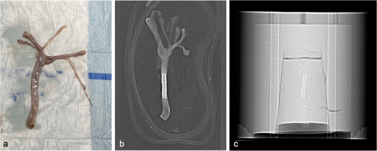
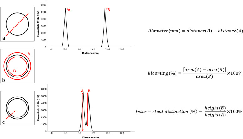
An ex vivo phantom was created, consisting of a 2% agar-water mixture, in which human-resected and stented arteries were individually embedded. Using similar technique parameters, helical scan data was acquired using a novel prototype Si-PCCT and a conventional EIDCT system at a volumetric CT dose index (CTDI) of 9 mGy. Reconstructions were made at 50 and 150 mm field-of-views (FOVs) using a bone kernel and adaptive statistical iterative reconstruction with 0% blending. Using a 5-point Likert scale, reader evaluations were performed on stent appearance, blooming and inter-stent visibility. Quantitative image analysis was performed on stent diameter accuracy, blooming and inter-stent distinction. Qualitative and quantitative differences between Si-PCCT and EIDCT systems were tested with a Wilcoxon signed-rank test and a paired samples t-test, respectively. Inter- and intra-reader agreement was assessed using the intraclass correlation coefficient (ICC).
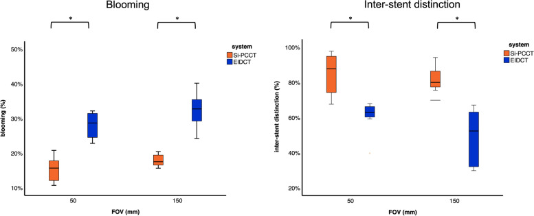
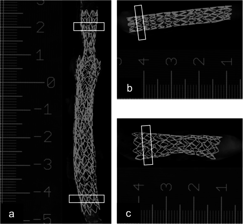
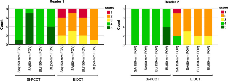
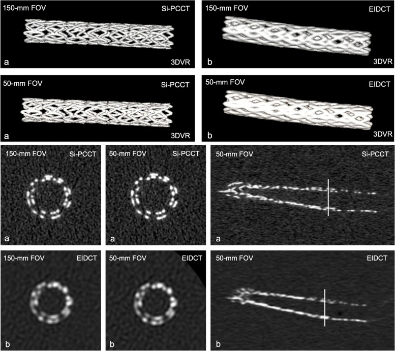
Qualitatively, Si-PCCT images were rated higher than EIDCT images at 150-mm FOV, based on stent appearance (p = 0.026) and blooming (p = 0.015), with a moderate inter- (ICC = 0.50) and intra-reader (ICC = 0.60) agreement. Quantitatively, Si-PCCT yielded more accurate diameter measurements (p = 0.001), reduced blooming (p < 0.001) and improved inter-stent distinction (p < 0.001). Similar trends were observed for the images reconstructed at 50-mm FOV.
When compared to EIDCT, the improved spatial resolution of Si-PCCT yields enhanced stent appearance, more accurate diameter measurements, reduced blooming and improved inter-stent distinction.
• This study evaluated stent appearance in a novel silicon-based photon-counting computed tomography (Si-PCCT) prototype. • Compared to standard CT, Si-PCCT resulted in more accurate stent diameter measurements. • Si-PCCT also reduced blooming artefacts and improved inter-stent visibility.
本研究比较了新型硅基光子计数计算机断层扫描(Si-PCCT)原型机与常规能量积分探测器 CT(EIDCT)系统中支架的成像表现。
制作了一个包含 2%琼脂水混合物的离体血管模型,其中单独嵌入了人切除和支架置入的动脉。使用类似的技术参数,在容积 CT 剂量指数(CTDI)为 9mGy 的情况下,使用新型 Si-PCCT 原型机和常规 EIDCT 系统进行螺旋扫描数据采集。使用骨核和自适应统计迭代重建(0%混合),在 50mm 和 150mm 视野(FOV)下进行重建。采用 5 分李克特量表,由 2 名读者对支架外观、blooming 和支架间可视性进行评价。对支架直径准确性、blooming 和支架间区分进行定量图像分析。使用 Wilcoxon 符号秩检验和配对样本 t 检验分别对 Si-PCCT 和 EIDCT 系统之间的定性和定量差异进行检验。使用组内相关系数(ICC)评估读者间和读者内的一致性。
定性分析显示,在 150mm FOV 下,Si-PCCT 图像的支架外观(p=0.026)和 blooming(p=0.015)评分均高于 EIDCT 图像,且读者间(ICC=0.50)和读者内(ICC=0.60)的一致性均为中度。定量分析显示,Si-PCCT 可获得更准确的直径测量值(p=0.001),减少 blooming(p<0.001)并改善支架间的区分(p<0.001)。在 50mm FOV 下,也观察到类似的趋势。
与 EIDCT 相比,Si-PCCT 的空间分辨率提高,从而改善了支架的成像外观,提高了支架直径测量的准确性,减少了 blooming 伪影,改善了支架间的区分。
•本研究评估了新型硅基光子计数计算机断层扫描(Si-PCCT)原型机中支架的成像表现。•与标准 CT 相比,Si-PCCT 可获得更准确的支架直径测量值。•Si-PCCT 还减少了 blooming 伪影,改善了支架间的可视性。