Department of Orthopaedics, Daping Hospital, Army Medical University, 10 Changjiang Branch Road, Yuzhong District, Chongqing, 400042, People's Republic of China.
Department of Orthopaedics, Chongqing Public Health Medical Center, Chongqing, 400030, People's Republic of China.
BMC Musculoskelet Disord. 2023 Apr 25;24(1):325. doi: 10.1186/s12891-023-06425-7.
To explore the mechanism of the healing of tendon tissue and anti-adhesion, and to discuss the role of the transforming growth factor-β3 (TGF-β3)/cAMP response element binding protein-1 (CREB-1) signaling pathway in the healing process of tendons.
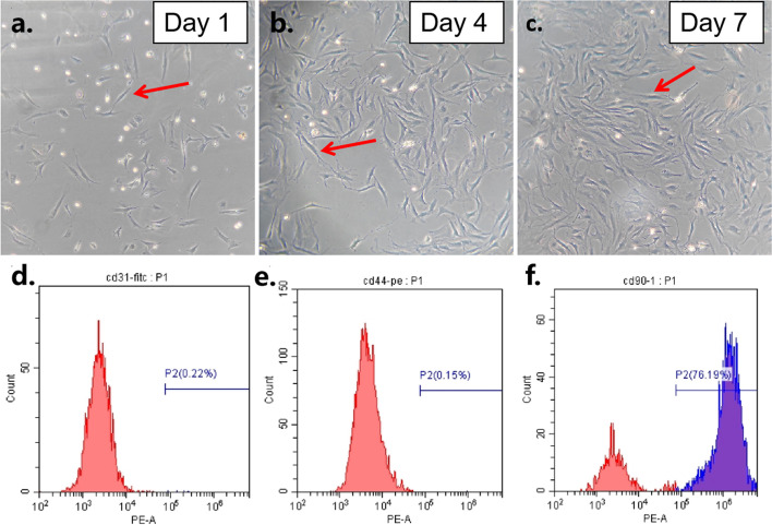
All mice were divided into four groups of 1, 2, 4, and 8 weeks respectively. Each time group was divided into four treatment groups: the amplification group, the inhibition group, the negative group, and the control group. When the tendon injury model was established, the CREB-1 virus was injected into the tendon injury parts. A series of methods such as gait behaviourism, anatomy, histological examination, immunohistochemical examination and collagen staining were employed to assess the tendon healing and the protein expression of TGF-β3, CREB-1, Smad3/7 and type I/III collagen (COL-I/III). CREB-1 virus was sent to tendon stem cells to assess the protein expression of TGF-β1, TGF-β3, CREB-1, COL-I/III by methods such as immunohistochemistry and Western blot.
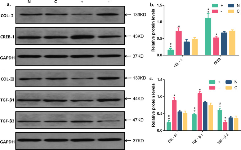
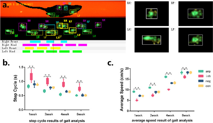
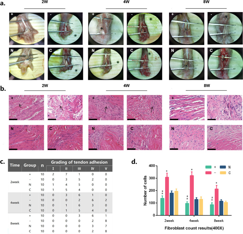
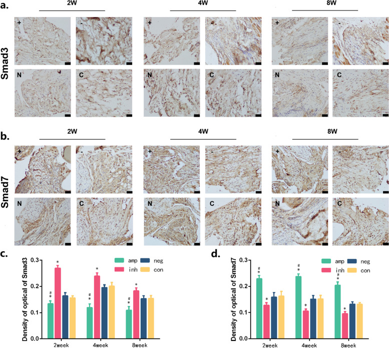
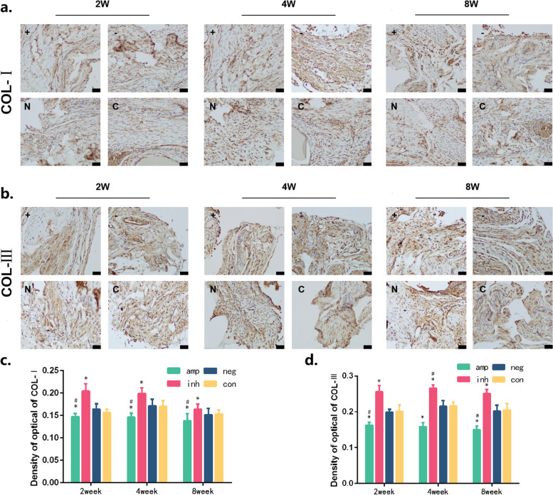
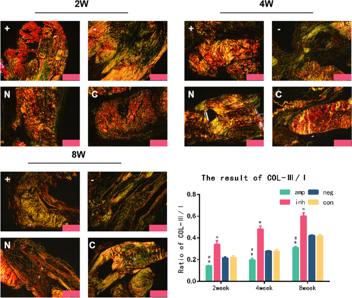
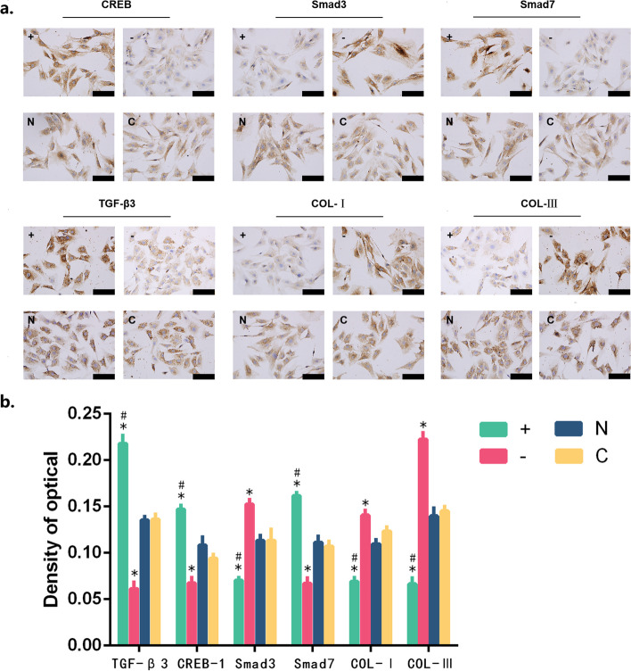
The amplification group showed better gait behaviourism than the inhibition group in the healing process. The amplification group also had less adhesion than the negative group. Hematoxylin-eosin (HE) staining of tendon tissue sections showed that the number of fibroblasts in the amplification group was less than the inhibition group, and the immunohistochemical results indicated that the expression of TGF-β3, CREB-1, and Smad7 at each time point was higher than the inhibition group. The expression of COL-I/III and Smad3 in the amplification group was lower than the inhibition group at all time points. The collagen staining indicated that the ratio of type I/III collagen in the amplification group was higher than the negative group at 2,4,8 week. The CREB-1 amplification virus could promote the protein expression of TGF-β3, CREB-1 and inhibit the protein expression of TGF-β1 and COL-I/III in the tendon stem cells.
In the process of tendon injury healing, CREB-1 could promote the secretion of TGF-β3, so as to promote the tendon healing and have the effect of anti-adhesion in tendons. It might provide new intervention targets for anti-adhesion treatment of tendon injuries.
探讨肌腱组织愈合和抗粘连的机制,探讨转化生长因子-β3(TGF-β3)/环磷酸腺苷反应元件结合蛋白-1(CREB-1)信号通路在肌腱愈合过程中的作用。
所有小鼠均分为 1、2、4、8 周 4 组,每组再分为扩增组、抑制组、阴性组和对照组。构建肌腱损伤模型后,将 CREB-1 病毒注入肌腱损伤部位。采用步态行为学、解剖学、组织学检查、免疫组织化学检查和胶原染色等方法评估肌腱愈合情况以及 TGF-β3、CREB-1、Smad3/7 和 I/III 型胶原(COL-I/III)的蛋白表达。将 CREB-1 病毒转染至肌腱干细胞,采用免疫组织化学和 Western blot 等方法检测 TGF-β1、TGF-β3、CREB-1、COL-I/III 的蛋白表达。
在愈合过程中,扩增组的步态行为学优于抑制组,且与阴性组相比,其粘连程度较轻。肌腱组织切片苏木精-伊红(HE)染色显示,扩增组的成纤维细胞数量少于抑制组,免疫组织化学结果表明,各时间点 TGF-β3、CREB-1 和 Smad7 的表达均高于抑制组。各时间点扩增组 COL-I/III 和 Smad3 的表达均低于抑制组。胶原染色显示,2、4、8 周时,扩增组 I/III 型胶原比例高于阴性组。CREB-1 扩增病毒可促进肌腱干细胞中 TGF-β3、CREB-1 蛋白的表达,抑制 TGF-β1 和 COL-I/III 蛋白的表达。
在肌腱损伤愈合过程中,CREB-1 可促进 TGF-β3 的分泌,从而促进肌腱愈合,在肌腱中具有抗粘连作用,为肌腱损伤抗粘连治疗提供新的干预靶点。