Lu Qifang, Chen Jin, Wang Yanming, Huang Li, Jiang Zhoufan, Nguchu Benedictor Alexander, Chen Shishuo, Qiu Bensheng, Wang Xiaoxiao
School of Clinical Medicine, Anhui Medical College, Anhui, China.
Center for Biomedical Imaging, University of Science and Technology of China, Anhui, China.
Psychiatry Investig. 2023 Apr;20(4):334-340. doi: 10.30773/pi.2022.0254. Epub 2023 Apr 20.
This study uses structural magnetic resonance imaging to explore changes in the cerebellar lobules in patients with autism spectrum disorder (ASD) and further analyze the correlation between cerebellar structural changes and clinical symptoms of ASD.
A total of 75 patients with ASD and 97 typically developing (TD) subjects from Autism Brain Imaging Data Exchange dataset were recruited. We adopted an advanced automatic cerebellar lobule segmentation technique called CEREbellum Segmentation to segment each cerebellar hemisphere into 12 lobules. Normalized cortical thickness of each lobule was recorded, and group differences in the cortical measures were evaluated. Correlation analysis was also performed between the normalized cortical thickness and the score of Autism Diagnostic Interview-Revised.
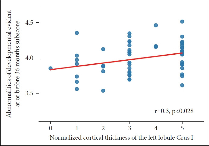
Results from analysis of variance showed that the normalized cortical thickness of the ASD group differed significantly from that of the TD group; specifically, the ASD group had lower normalized cortical thickness than the TD group. Post-hoc analysis revealed that the differences were more predominant in the left lobule VI, left lobule Crus I and left lobule X, and in the right lobule VI and right lobule Crus I. Lowered normalized cortical thickness in the left lobule Crus I in the ASD patients correlated positively with the abnormality of development evident at or before 36 months subscore.
These results suggest abnormal development of cerebellar lobule structures in ASD patients, and such abnormality might significantly influence the pathogenesis of ASD. These findings provide new insights into the neural mechanisms of ASD, which may be clinically relevant to ASD diagnosis.
本研究采用结构磁共振成像技术,探究自闭症谱系障碍(ASD)患者小脑小叶的变化,并进一步分析小脑结构变化与ASD临床症状之间的相关性。
从自闭症脑成像数据交换数据集中招募了75例ASD患者和97名发育正常(TD)的受试者。我们采用一种先进的自动小脑小叶分割技术,即小脑分割技术,将每个小脑半球分割为12个小叶。记录每个小叶的标准化皮质厚度,并评估两组在皮质测量指标上的差异。同时,还对标准化皮质厚度与自闭症诊断访谈修订版的得分进行了相关性分析。
方差分析结果显示,ASD组的标准化皮质厚度与TD组存在显著差异;具体而言,ASD组的标准化皮质厚度低于TD组。事后分析表明,差异在左侧小叶VI、左侧小叶 Crus I和左侧小叶X以及右侧小叶VI和右侧小叶 Crus I中更为明显。ASD患者左侧小叶 Crus I的标准化皮质厚度降低与36个月及以前发育异常子得分呈正相关。
这些结果表明ASD患者小脑小叶结构发育异常,这种异常可能对ASD的发病机制产生显著影响。这些发现为ASD的神经机制提供了新的见解,可能与ASD的临床诊断相关。