The Hormel Institute, University of Minnesota.
The Hormel Institute, University of Minnesota;
J Vis Exp. 2023 Apr 21(194). doi: 10.3791/65118.
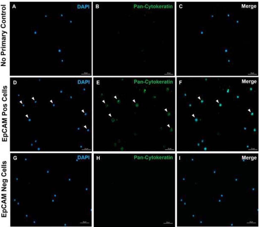
Epithelial cells have been identified in the blood and bone marrow of patients with cancer and other diseases. However, the presence of normal epithelial cells in the blood and bone marrow of healthy individuals has yet to be identified in a consistent way. Presented here is a reproducible method for isolating epithelial cells from healthy human and murine blood and bone marrow (BM) using flow cytometry and immunofluorescence (IF) microscopy. Epithelial cells in healthy individuals were first identified and isolated via flow cytometry using epithelial cell adhesion molecule (EpCAM). These EpCAM+ cells were confirmed to express keratin using immunofluorescence microscopy in Krt1-14;mTmG transgenic mice. Human blood samples had 0.18% ± 0.0004 EpCAM+ cells (SEM; n=7 biological replicates, 4 experimental replicates). In human BM, 3.53% ± 0.006 (SEM; n=3 biological replicates, 4 experimental replicates) of mononuclear cells were EpCAM+. In mouse blood, EpCAM+ cells constituted 0.45% ± 0.0006 (SEM; n=2 biological replicates, 4 experimental replicates), and in mouse BM, 5.17% ± 0.001 (SEM; n=3 biological replicates, 4 experimental replicates) were EpCAM+. In mice, all the EpCAM+ cells were immunoreactive to pan-cytokeratin, as determined by IF microscopy. Results were confirmed using Krt1-14;mTmG transgenic mice, with low (8.6 native GFP+ cells per 10 cells analyzed; 0.085% of viable cells), but significant numbers (p < 0.0005) of GFP+ cells present in normal murine BM, that were not the result of randomness compared with multiple negative controls. Further, EpCAM+ cells in mouse blood were more heterogeneous than CD45+ cells (0.58% in BM; 0.13% in blood). These observations conclude that cells expressing cytokeratin proteins are reproducibly detectable among mononuclear cells from human and murine blood and BM. We demonstrate a method of tissue harvesting, flow cytometry, and immunostaining that can be used to identify and determine the function of these pan-cytokeratin epithelial cells in healthy individuals.
已经在癌症和其他疾病患者的血液和骨髓中鉴定出上皮细胞。然而,在健康个体的血液和骨髓中,尚未以一致的方式鉴定出正常上皮细胞的存在。本文介绍了一种从健康人及鼠的血液和骨髓(BM)中分离上皮细胞的可重复方法,该方法使用流式细胞术和免疫荧光(IF)显微镜。首先,通过流式细胞术使用上皮细胞黏附分子(EpCAM)鉴定并分离健康个体中的上皮细胞。在 Krt1-14;mTmG 转基因小鼠中,通过免疫荧光显微镜证实这些 EpCAM+细胞表达角蛋白。人血样本中 EpCAM+细胞的含量为 0.18%±0.0004(SEM;n=7 个生物学重复,4 个实验重复)。在人 BM 中,单核细胞中有 3.53%±0.006(SEM;n=3 个生物学重复,4 个实验重复)为 EpCAM+。在鼠血中,EpCAM+细胞构成 0.45%±0.0006(SEM;n=2 个生物学重复,4 个实验重复),而在鼠 BM 中,5.17%±0.001(SEM;n=3 个生物学重复,4 个实验重复)为 EpCAM+。在小鼠中,所有 EpCAM+细胞均通过 IF 显微镜鉴定为泛细胞角蛋白阳性。结果通过 Krt1-14;mTmG 转基因小鼠得到了验证,该小鼠的正常鼠 BM 中存在低水平(每 10 个分析细胞中有 8.6 个天然 GFP+细胞;占活细胞的 0.085%)但具有统计学意义(p<0.0005)的 GFP+细胞,与多个阴性对照相比,这些 GFP+细胞并非随机存在。此外,鼠血中的 EpCAM+细胞比 CD45+细胞更具异质性(BM 中为 0.58%;血液中为 0.13%)。这些观察结果表明,在人及鼠的血液和 BM 中的单核细胞中,可重复检测到表达细胞角蛋白蛋白的细胞。我们展示了一种组织采集、流式细胞术和免疫染色的方法,可用于鉴定和确定这些健康个体中泛细胞角蛋白上皮细胞的功能。