Laboratory for Equine and Comparative Orthopedic Research, Department of Veterinary Clinical Sciences, School of Veterinary Medicine, Louisiana State University, Baton Rouge, Louisiana, United States of America.
PLoS One. 2023 May 8;18(5):e0285475. doi: 10.1371/journal.pone.0285475. eCollection 2023.
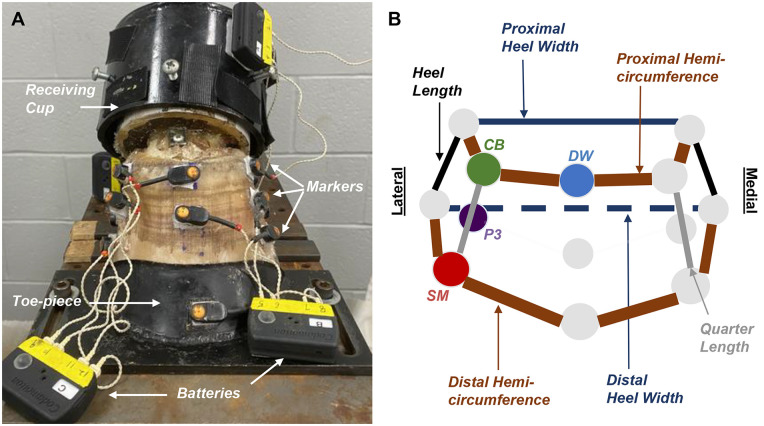
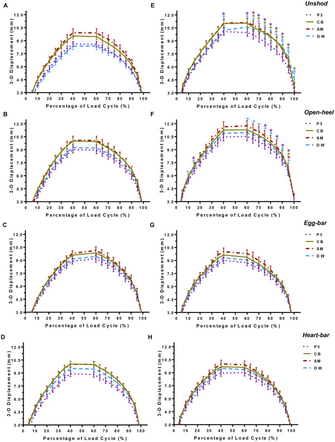
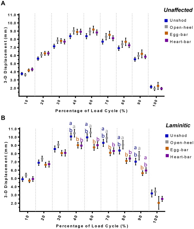
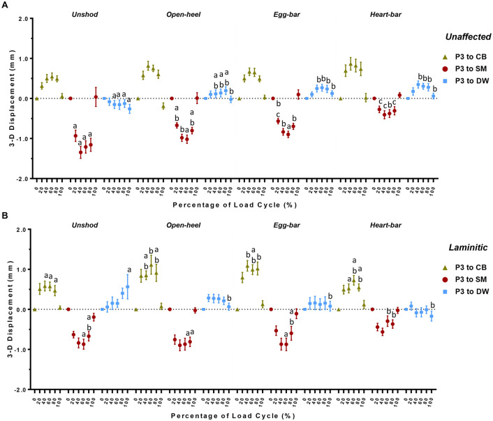
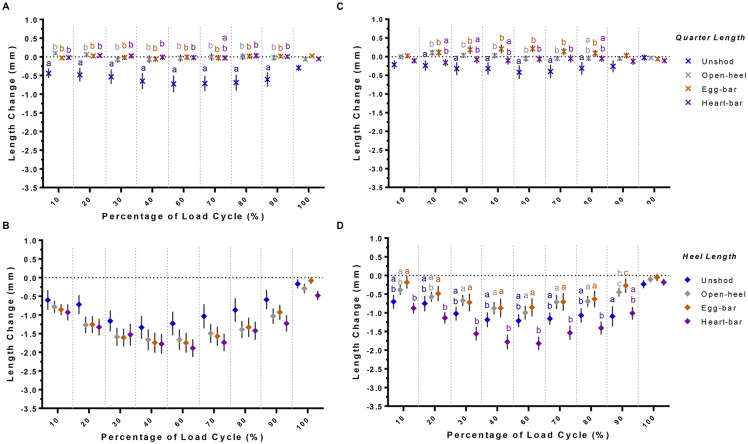
Equine shoes provide hoof protection and support weakened or damaged hoof tissues. Two hypotheses were tested in this study: 1) motion of the third phalanx (P3) and hoof wall deformation are greater in laminitic versus unaffected hooves regardless of shoe type; 2) P3 displacement and hoof wall deformation are greatest while unshod (US), less with open-heel (OH), then egg-bar (EB) shoes, and least with heart-bar (HB) shoes for both hoof conditions. Distal forelimbs (8/condition) were subjected to compressive forces (1.0x10²-5.5x10³ N) while a real-time motion detection system recorded markers on P3 and the hoof wall coronary band, vertical midpoint, and solar margin. Magnitude and direction of P3 displacement and changes in proximal and distal hemi-circumference, quarter and heel height and proximal and distal heel width were quantified. Hoof condition and shoe effects were assessed with 2-way ANOVA (p<0.05). P3 displacement was greater in laminitic hooves when US or with OH, and EB and HB reduced P3 displacement in laminitic hooves. P3 displacement was similar among shoes in unaffected hooves and greatest in laminitic hooves with OH, then US, EB and HB. EB and HB increased P3 displacement from the dorsal wall in unaffected hooves and decreased it in laminitic hooves. OH and EB increased P3 motion from the coronary band in laminitic hooves, and HB decreased P3 motion toward the solar margin in unaffected and laminitic hooves. In laminitic hooves, HB reduced distal hemi-circumference and quarter deformation and increased heel deformation and expansion. Proximal hemi-circumference constriction was inversely related to proximal heel expansion with and without shoes. Overall, shoe configuration alters hoof deformation distinctly between unaffected and laminitic hooves, and HB provided the greatest P3 stability in laminitic hooves. These unique results about P3 motion and hoof deformation in laminitic and unaffected hooves inform shoe selection and design.
马掌为马蹄提供保护,并支撑虚弱或受损的蹄组织。本研究检验了两个假设:1)无论鞋型如何,蹄叶炎马的第三跖骨(P3)运动和蹄壁变形都大于正常马;2)在未穿鞋(US)时,P3 位移和蹄壁变形最大,开放式鞋(OH)次之,蹄铁(EB)最小,蹄叶炎马和正常马的蹄心铁(HB)最小。将 8 条前肢(每条条件)置于压缩力(1.0x10²-5.5x10³ N)下,同时实时运动检测系统记录 P3 和蹄壁冠状带、垂直中点、太阳缘上的标记。P3 位移的大小和方向,以及近端和远端半周长、 quarters 和 heel height 以及近端和远端 heel width 的变化均进行了量化。采用 2 因素方差分析(p<0.05)评估蹄状况和鞋的影响。US 或 OH 时,蹄叶炎马的 P3 位移更大,而 EB 和 HB 则减少了蹄叶炎马的 P3 位移。正常马的各鞋型 P3 位移相似,而 OH 时蹄叶炎马的 P3 位移最大,其次是 US、EB 和 HB。EB 和 HB 增加了正常马蹄壁的 P3 位移,减少了蹄叶炎马的 P3 位移。OH 和 EB 增加了蹄叶炎马冠状带的 P3 运动,HB 减少了正常和蹄叶炎马向太阳缘的 P3 运动。在蹄叶炎马中,HB 减少了远端半周长和 quarters 的变形,增加了 heel 的变形和扩张。近端半周长的收缩与有或无鞋时近端 heel 的扩张呈反比。总的来说,鞋型配置明显改变了正常和蹄叶炎马蹄的蹄变形,而 HB 在蹄叶炎马中提供了最大的 P3 稳定性。这些关于蹄叶炎和正常马蹄 P3 运动和蹄变形的独特结果为鞋的选择和设计提供了信息。