• 文献检索
  • 文档翻译
  • 深度研究
  • 学术资讯
  • Suppr Zotero 插件Zotero 插件
  • 邀请有礼
  • 套餐&价格
  • 历史记录
应用&插件
Suppr Zotero 插件Zotero 插件浏览器插件Mac 客户端Windows 客户端微信小程序
定价
高级版会员购买积分包购买API积分包
服务
文献检索文档翻译深度研究API 文档MCP 服务
关于我们
关于 Suppr公司介绍联系我们用户协议隐私条款
关注我们

Suppr 超能文献

核心技术专利:CN118964589B侵权必究
粤ICP备2023148730 号-1Suppr @ 2026

文献检索

告别复杂PubMed语法,用中文像聊天一样搜索,搜遍4000万医学文献。AI智能推荐,让科研检索更轻松。

立即免费搜索

文件翻译

保留排版,准确专业,支持PDF/Word/PPT等文件格式,支持 12+语言互译。

免费翻译文档

深度研究

AI帮你快速写综述,25分钟生成高质量综述,智能提取关键信息,辅助科研写作。

立即免费体验

优化 4D 流 MRI 的空间和时间分辨率,以检查颈动脉分叉处的复杂血液动力学。

Optimization of 4D Flow MRI Spatial and Temporal Resolution for Examining Complex Hemodynamics in the Carotid Artery Bifurcation.

机构信息

Department of Biomedical Engineering, The Wallace H. Coulter, Emory University and Georgia Institute of Technology, Atlanta, GA, USA.

Department of Radiology & Imaging Sciences, Emory University, 1364 Clifton Rd, Atlanta, GA, 30322, USA.

出版信息

Cardiovasc Eng Technol. 2023 Jun;14(3):476-488. doi: 10.1007/s13239-023-00667-1. Epub 2023 May 8.

DOI:10.1007/s13239-023-00667-1
PMID:37156900
原文链接:https://pmc.ncbi.nlm.nih.gov/articles/PMC10524741/
Abstract

BACKGROUND

Three-dimensional, ECG-gated, time-resolved, three-directional, velocity-encoded phase-contrast MRI (4D flow MRI) has been applied extensively to measure blood velocity in great vessels but has been much less used in diseased carotid arteries. Carotid artery webs (CaW) are non-inflammatory intraluminal shelf-like projections into the internal carotid artery (ICA) bulb that are associated with complex flow and cryptogenic stroke.

PURPOSE

Optimize 4D flow MRI for measuring the velocity field of complex flow in the carotid artery bifurcation model that contains a CaW.

METHODS

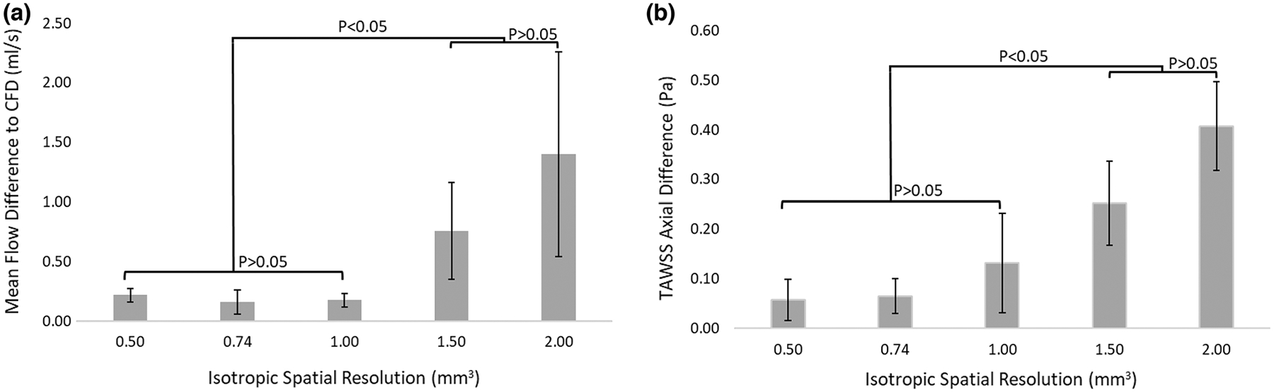
A 3D printed phantom model created from computed tomography angiography (CTA) of a subject with CaW was placed in a pulsatile flow loop within the MRI scanner. 4D Flow MRI images of the phantom were acquired with five different spatial resolutions (0.50-2.00  mm) and four different temporal resolutions (23-96 ms) and compared to a computational fluid dynamics (CFD) solution of the flow field as a reference. We examined four planes perpendicular to the vessel centerline, one in the common carotid artery (CCA) and three in the internal carotid artery (ICA) where complex flow was expected. At these four planes pixel-by-pixel velocity values, flow, and time average wall shear stress (TAWSS) were compared between 4D flow MRI and CFD.

HYPOTHESIS

An optimized 4D flow MRI protocol will provide a good correlation with CFD velocity and TAWSS values in areas of complex flow within a clinically feasible scan time (~ 10 min).

RESULTS

Spatial resolution affected the velocity values, time average flow, and TAWSS measurements. Qualitatively, a spatial resolution of 0.50  mm resulted in higher noise, while a lower spatial resolution of 1.50-2.00  mm did not adequately resolve the velocity profile. Isotropic spatial resolutions of 0.50-1.00  mm showed no significant difference in total flow compared to CFD. Pixel-by-pixel velocity correlation coefficients between 4D flow MRI and CFD were > 0.75 for 0.50-1.00  mm but were < 0.5 for 1.50 and 2.00  mm. Regional TAWSS values determined from 4D flow MRI were generally lower than CFD and decreased at lower spatial resolutions (larger pixel sizes). TAWSS differences between 4D flow and CFD were not statistically significant at spatial resolutions of 0.50-1.00  mm but were different at 1.50 and 2.00 mm. Differences in temporal resolution only affected the flow values when temporal resolution was > 48.4 ms; temporal resolution did not affect TAWSS values.

CONCLUSION

A spatial resolution of 0.74-1.00  mm and a temporal resolution of 23-48 ms (1-2 k-space segments) provides a 4D flow MRI protocol capable of imaging velocity and TAWSS in regions of complex flow within the carotid bifurcation at a clinically acceptable scan time.

摘要

背景

三维、心电图门控、时分辨、三向、速度编码相位对比磁共振成像(4D 流 MRI)已广泛应用于测量大动脉中的血流速度,但在病变颈动脉中的应用较少。颈动脉网(CaW)是向颈内动脉(ICA)球内突起的非炎症性腔内架状突起,与复杂的血流和隐源性中风有关。

目的

优化 4D 流 MRI,以测量包含 CaW 的颈动脉分叉模型中的复杂血流速度场。

方法

使用来自伴有 CaW 的患者的 CT 血管造影(CTA)创建的 3D 打印体模,将其放置在 MRI 扫描仪内的脉动流回路中。使用五个不同的空间分辨率(0.50-2.00 毫米)和四个不同的时间分辨率(23-96 毫秒)获取体模的 4D 流 MRI 图像,并与作为参考的流场计算流体动力学(CFD)解进行比较。我们检查了与血管中心线垂直的四个平面,一个在颈总动脉(CCA),三个在颈内动脉(ICA),预计在这些平面上会出现复杂的血流。在这四个平面上,逐像素比较 4D 流 MRI 和 CFD 的速度值、流量和时间平均壁切应力(TAWSS)。

假设

在临床可行的扫描时间(~10 分钟)内,优化的 4D 流 MRI 协议将与 CFD 速度和 TAWSS 值具有良好的相关性,适用于复杂血流区域。

结果

空间分辨率会影响速度值、时间平均流量和 TAWSS 测量值。定性地说,0.50 毫米的空间分辨率会导致更高的噪声,而较低的 1.50-2.00 毫米的空间分辨率则不能充分分辨速度分布。0.50-1.00 毫米各向同性空间分辨率与 CFD 相比,总流量无显著差异。4D 流 MRI 和 CFD 之间的逐像素速度相关系数大于 0.75 毫米,但小于 0.5 毫米 1.50 和 2.00 毫米。4D 流 MRI 确定的局部 TAWSS 值通常低于 CFD,并且随着空间分辨率(较大的像素尺寸)的降低而降低。在 0.50-1.00 毫米的空间分辨率下,4D 流和 CFD 之间的 TAWSS 差异没有统计学意义,但在 1.50 和 2.00 毫米时则不同。时间分辨率的差异仅在时间分辨率大于 48.4 毫秒时才会影响流量值;时间分辨率不会影响 TAWSS 值。

结论

空间分辨率为 0.74-1.00 毫米,时间分辨率为 23-48 毫秒(1-2 个 k-空间段),可在临床可接受的扫描时间内对颈动脉分叉处的复杂血流区域进行成像,提供 4D 流 MRI 协议,以测量速度和 TAWSS。

https://cdn.ncbi.nlm.nih.gov/pmc/blobs/498b/10524741/041d833e6417/nihms-1901196-f0007.jpg
https://cdn.ncbi.nlm.nih.gov/pmc/blobs/498b/10524741/feb041d71e1e/nihms-1901196-f0001.jpg
https://cdn.ncbi.nlm.nih.gov/pmc/blobs/498b/10524741/df9f0496eb71/nihms-1901196-f0002.jpg
https://cdn.ncbi.nlm.nih.gov/pmc/blobs/498b/10524741/ea6c25ae5971/nihms-1901196-f0003.jpg
https://cdn.ncbi.nlm.nih.gov/pmc/blobs/498b/10524741/2807406b2ac0/nihms-1901196-f0004.jpg
https://cdn.ncbi.nlm.nih.gov/pmc/blobs/498b/10524741/75840f2449d6/nihms-1901196-f0005.jpg
https://cdn.ncbi.nlm.nih.gov/pmc/blobs/498b/10524741/eb781ac39956/nihms-1901196-f0006.jpg
https://cdn.ncbi.nlm.nih.gov/pmc/blobs/498b/10524741/041d833e6417/nihms-1901196-f0007.jpg

相似文献

1
Optimization of 4D Flow MRI Spatial and Temporal Resolution for Examining Complex Hemodynamics in the Carotid Artery Bifurcation.优化 4D 流 MRI 的空间和时间分辨率,以检查颈动脉分叉处的复杂血液动力学。
Cardiovasc Eng Technol. 2023 Jun;14(3):476-488. doi: 10.1007/s13239-023-00667-1. Epub 2023 May 8.
2
Assessment of Complex Flow Patterns in Patients With Carotid Webs, Patients With Carotid Atherosclerosis, and Healthy Subjects Using 4D Flow MRI.应用 4D 血流 MRI 评估颈动脉瘤患者、颈动脉粥样硬化患者和健康受试者的复杂血流模式。
J Magn Reson Imaging. 2024 Jun;59(6):2001-2010. doi: 10.1002/jmri.29013. Epub 2023 Sep 14.
3
The impact of 4D-Flow MRI spatial resolution on patient-specific CFD simulations of the thoracic aorta.4D-Flow MRI 空间分辨率对胸主动脉患者特定 CFD 模拟的影响。
Sci Rep. 2022 Sep 6;12(1):15128. doi: 10.1038/s41598-022-19347-6.
4
Four-Dimensional Flow Magnetic Resonance Imaging for Assessment of Velocity Magnitudes and Flow Patterns in The Human Carotid Artery Bifurcation: Comparison with Computational Fluid Dynamics.用于评估人体颈动脉分叉处速度大小和血流模式的四维流动磁共振成像:与计算流体动力学的比较
Diagnostics (Basel). 2019 Dec 13;9(4):223. doi: 10.3390/diagnostics9040223.
5
Assessment of hepatic arterial hemodynamics with 4D flow MRI: in vitro analysis of motion and spatial resolution related error and in vivo feasibility study in 20 volunteers.应用 4D 血流 MRI 评估肝动脉血流动力学:运动和空间分辨率相关误差的离体分析及 20 例志愿者的体内可行性研究。
Eur Radiol. 2022 Dec;32(12):8639-8648. doi: 10.1007/s00330-022-08890-5. Epub 2022 Jun 22.
6
Hemodynamics of human carotid artery bifurcations: computational studies with models reconstructed from magnetic resonance imaging of normal subjects.人体颈动脉分叉处的血流动力学:基于正常受试者磁共振成像重建模型的计算研究
J Vasc Surg. 1998 Jul;28(1):143-56. doi: 10.1016/s0741-5214(98)70210-1.
7
Age and anatomical location related hemodynamic changes assessed by 4D flow MRI in the carotid arteries of healthy adults.4D 流 MRI 评估健康成年人颈动脉的年龄和解剖位置相关血流动力学变化。
Eur J Radiol. 2020 Jul;128:109035. doi: 10.1016/j.ejrad.2020.109035. Epub 2020 May 4.
8
Comparison of Prospective and Retrospective Gated 4D Flow Cardiac MR Image Acquisitions in the Carotid Bifurcation.前瞻性和回顾性门控 4D 流心脏磁共振成像在颈动脉分叉处的比较。
Cardiovasc Eng Technol. 2023 Feb;14(1):1-12. doi: 10.1007/s13239-022-00630-6. Epub 2022 May 26.
9
Variability of 4D flow parameters when subjected to changes in MRI acquisition parameters using a realistic thoracic aortic phantom.使用真实的胸主动脉体模研究 MRI 采集参数变化时 4D 流参数的可变性。
Magn Reson Med. 2018 Apr;79(4):1882-1892. doi: 10.1002/mrm.26834. Epub 2017 Jul 16.
10
Peak velocity measurements in tortuous arteries with phase contrast magnetic resonance imaging: the effect of multidirectional velocity encoding.迂曲动脉内峰值速度测量:相位对比磁共振成像中多方向速度编码的影响。
Invest Radiol. 2014 Apr;49(4):189-94. doi: 10.1097/RLI.0000000000000013.

引用本文的文献

1
Observation of intracranial artery and venous sinus hemodynamics using compressed sensing-accelerated 4D flow MRI: performance at different acceleration factors.使用压缩感知加速4D血流MRI观察颅内动脉和静脉窦血流动力学:不同加速因子下的性能表现
Front Neurosci. 2024 Jul 25;18:1438003. doi: 10.3389/fnins.2024.1438003. eCollection 2024.
2
The hemodynamic and geometric characteristics of carotid artery atherosclerotic plaque formation.颈动脉粥样硬化斑块形成的血流动力学和几何特征。
Quant Imaging Med Surg. 2024 Jul 1;14(7):4348-4361. doi: 10.21037/qims-23-1827. Epub 2024 Jun 11.
3
Subjects with carotid webs demonstrate pro-thrombotic hemodynamics compared to subjects with carotid atherosclerosis.

本文引用的文献

1
Particle Image Velocimetry of 3D-Printed Anatomical Blood Vascular Models Affected by Atherosclerosis.受动脉粥样硬化影响的3D打印解剖学血管模型的粒子图像测速技术
Materials (Basel). 2023 Jan 25;16(3):1055. doi: 10.3390/ma16031055.
2
A New Method for Quantifying Abdominal Aortic Wall Shear Stress Using Phase Contrast Magnetic Resonance Imaging and the Womersley Solution.一种使用相位对比磁共振成像和沃默斯利解定量计算腹主动脉壁切应力的新方法。
J Biomech Eng. 2022 Sep 1;144(9). doi: 10.1115/1.4054236.
3
In Vitro Validation of Regional Circumferential Strain Assessment in a Phantom Aortic Model Using Cine Displacement Encoding With Stimulated Echoes MRI.
与颈动脉粥样硬化患者相比,颈动脉膜患者表现出促血栓形成的血液动力学特征。
Sci Rep. 2024 May 2;14(1):10092. doi: 10.1038/s41598-024-60666-7.
4
Assessment of Complex Flow Patterns in Patients With Carotid Webs, Patients With Carotid Atherosclerosis, and Healthy Subjects Using 4D Flow MRI.应用 4D 血流 MRI 评估颈动脉瘤患者、颈动脉粥样硬化患者和健康受试者的复杂血流模式。
J Magn Reson Imaging. 2024 Jun;59(6):2001-2010. doi: 10.1002/jmri.29013. Epub 2023 Sep 14.
使用磁共振成像的电影位移编码与激发回波技术在模拟主动脉模型中对局部圆周应变评估的体外验证
J Magn Reson Imaging. 2022 Jun;55(6):1773-1784. doi: 10.1002/jmri.27972. Epub 2021 Oct 27.
4
Improving Blood Flow Visualization of Recirculation Regions at Carotid Bulb in 4D Flow MRI Using Semi-Automatic Segmentation with ITK-SNAP.使用ITK-SNAP半自动分割技术改善4D流磁共振成像中颈动脉球部再循环区域的血流可视化
Diagnostics (Basel). 2021 Oct 13;11(10):1890. doi: 10.3390/diagnostics11101890.
5
Turbulence Intensity as an Indicator for Ischemic Stroke in the Carotid Web.颈动脉窦湍流强度作为缺血性卒中的一个指标。
World Neurosurg. 2021 Oct;154:e443-e457. doi: 10.1016/j.wneu.2021.07.049. Epub 2021 Jul 26.
6
Carotid webs produce greater hemodynamic disturbances than atherosclerotic disease: a DSA time-density curve study.颈动脉梳状结构比动脉粥样硬化性疾病产生更大的血液动力学紊乱:一项 DSA 时间密度曲线研究。
J Neurointerv Surg. 2022 Jul;14(7):729-733. doi: 10.1136/neurintsurg-2021-017588. Epub 2021 Jul 27.
7
Carotid web leads to new thrombus formation: computational fluid dynamic analysis coupled with histological evidence.颈动脉壁梳状结构导致新血栓形成:计算流体动力学分析结合组织学证据。
Acta Neurochir (Wien). 2020 Oct;162(10):2583-2588. doi: 10.1007/s00701-020-04272-2. Epub 2020 Mar 10.
8
Four-Dimensional Flow Magnetic Resonance Imaging for Assessment of Velocity Magnitudes and Flow Patterns in The Human Carotid Artery Bifurcation: Comparison with Computational Fluid Dynamics.用于评估人体颈动脉分叉处速度大小和血流模式的四维流动磁共振成像:与计算流体动力学的比较
Diagnostics (Basel). 2019 Dec 13;9(4):223. doi: 10.3390/diagnostics9040223.
9
Flow Patterns in Carotid Webs: A Patient-Based Computational Fluid Dynamics Study.颈动脉瘤内流型:基于患者的计算流体动力学研究。
AJNR Am J Neuroradiol. 2019 Apr;40(4):703-708. doi: 10.3174/ajnr.A6012. Epub 2019 Mar 14.
10
Reconciling PC-MRI and CFD: An in-vitro study.PC-MRI 与 CFD 融合:一项体外研究。
NMR Biomed. 2019 May;32(5):e4063. doi: 10.1002/nbm.4063. Epub 2019 Feb 12.