Choi Moon Hyung, Kim Dong Hwan, Lee Young Joon, Rha Sung Eun, Lee Ji Youl
Department of Radiology, Eunpyeong St. Mary's Hospital, College of Medicine, The Catholic University of Korea, Seoul, Republic of Korea.
Department of Radiology, Seoul St. Mary's Hospital, College of Medicine, The Catholic University of Korea, 222 Banpo-daero, Seocho-gu, Seoul, 06591, Republic of Korea.
Insights Imaging. 2023 May 8;14(1):77. doi: 10.1186/s13244-023-01422-9.
To systematically determine the diagnostic performance of each MRI feature of the PI-RADS for predicting extraprostatic extension (EPE) in prostate cancer.
A literature search in the MEDLINE and EMBASE databases was conducted to identify original studies reporting the accuracy of each feature on MRI for the dichotomous diagnosis of EPE. The meta-analytic pooled diagnostic odds ratio (DOR), sensitivity, specificity, and their 95% confidence intervals (CIs) were obtained using a bivariate random-effects model.
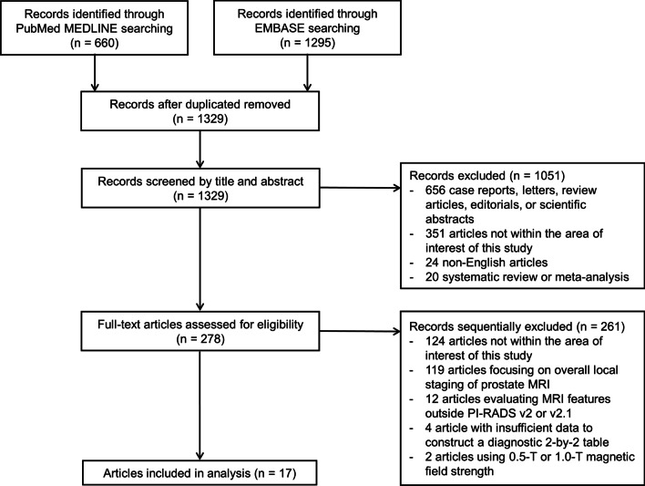
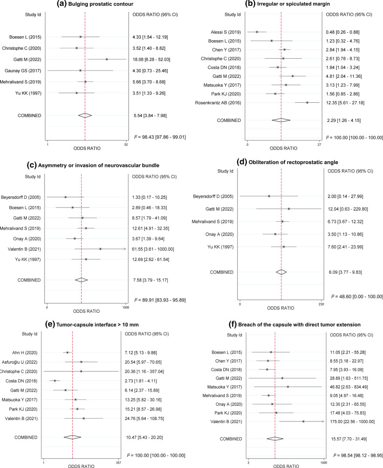
After screening 1955 studies, 17 studies with a total of 3062 men were included. All six imaging features, i.e., bulging prostatic contour, irregular or spiculated margin, asymmetry or invasion of neurovascular bundle, obliteration of rectoprostatic angle, tumor-capsule interface > 10 mm, and breach of the capsule with evidence of direct tumor extension, were significantly associated with EPE. Breach of the capsule with direct tumor extension demonstrated the highest pooled DOR (15.6, 95% CI [7.7-31.5]) followed by tumor-capsule interface > 10 mm (10.5 [5.4-20.2]), asymmetry or invasion of neurovascular bundle (7.6 [3.8-15.2]), and obliteration of rectoprostatic angle (6.1 [3.8-9.8]). Irregular or spiculated margin showed the lowest pooled DOR (2.3 [1.3-4.2]). Breach of the capsule with direct tumor extension and tumor-capsule interface > 10 mm showed the highest pooled specificity (98.0% [96.2-99.0]) and sensitivity (86.3% [70.0-94.4]), respectively.
Among the six MRI features of prostate cancer, breach of the capsule with direct tumor extension and tumor-capsule interface > 10 mm were the most predictive of EPE with the highest specificity and sensitivity, respectively.
系统地确定前列腺影像报告和数据系统(PI-RADS)的每个MRI特征对预测前列腺癌前列腺外侵犯(EPE)的诊断效能。
在MEDLINE和EMBASE数据库中进行文献检索,以识别报告MRI上每个特征对EPE进行二分诊断准确性的原始研究。使用双变量随机效应模型获得荟萃分析合并诊断比值比(DOR)、敏感性、特异性及其95%置信区间(CI)。
在筛选1955项研究后,纳入了17项研究,共3062名男性。所有六个影像特征,即前列腺轮廓凸出、边缘不规则或呈毛刺状、神经血管束不对称或受侵犯、直肠前列腺角消失、肿瘤-包膜界面>10mm以及包膜破裂且有肿瘤直接蔓延的证据,均与EPE显著相关。有肿瘤直接蔓延的包膜破裂显示合并DOR最高(15.6,95%CI[7.7-31.5]),其次是肿瘤-包膜界面>10mm(10.5[5.4-20.2])、神经血管束不对称或受侵犯(7.6[3.8-15.2])以及直肠前列腺角消失(6.1[3.8-9.8])。边缘不规则或呈毛刺状显示合并DOR最低(2.3[1.3-4.2])。有肿瘤直接蔓延的包膜破裂和肿瘤-包膜界面>10mm分别显示合并特异性最高(98.0%[96.2-99.0])和敏感性最高(86.3%[70.0-94.4])。
在前列腺癌的六个MRI特征中,有肿瘤直接蔓延的包膜破裂和肿瘤-包膜界面>10mm分别是EPE最具预测性的特征,特异性和敏感性最高。