• 文献检索
  • 文档翻译
  • 深度研究
  • 学术资讯
  • Suppr Zotero 插件Zotero 插件
  • 邀请有礼
  • 套餐&价格
  • 历史记录
应用&插件
Suppr Zotero 插件Zotero 插件浏览器插件Mac 客户端Windows 客户端微信小程序
定价
高级版会员购买积分包购买API积分包
服务
文献检索文档翻译深度研究API 文档MCP 服务
关于我们
关于 Suppr公司介绍联系我们用户协议隐私条款
关注我们

Suppr 超能文献

核心技术专利:CN118964589B侵权必究
粤ICP备2023148730 号-1Suppr @ 2026

文献检索

告别复杂PubMed语法,用中文像聊天一样搜索,搜遍4000万医学文献。AI智能推荐,让科研检索更轻松。

立即免费搜索

文件翻译

保留排版,准确专业,支持PDF/Word/PPT等文件格式,支持 12+语言互译。

免费翻译文档

深度研究

AI帮你快速写综述,25分钟生成高质量综述,智能提取关键信息,辅助科研写作。

立即免费体验

mEPE 评分:一种用于预测前列腺癌多参数磁共振成像下病理性前列腺外延伸的综合分级系统。

mEPE-score: a comprehensive grading system for predicting pathologic extraprostatic extension of prostate cancer at multiparametric magnetic resonance imaging.

机构信息

Department of Surgical Sciences, Radiology Unit, University of Turin, Via Genova 3, 10126, Turin, Italy.

Urology Unit, Department of Surgical Sciences, University of Turin, Turin, Italy.

出版信息

Eur Radiol. 2022 Jul;32(7):4942-4953. doi: 10.1007/s00330-022-08595-9. Epub 2022 Mar 15.

DOI:10.1007/s00330-022-08595-9
PMID:35290508
原文链接:https://pmc.ncbi.nlm.nih.gov/articles/PMC9213375/
Abstract

OBJECTIVE

To investigate the diagnostic accuracy of the PI-RADS v2.1 multiparametric magnetic resonance imaging (mpMRI) features in predicting extraprostatic extension (mEPE) of prostate cancer (PCa), as well as to develop and validate a comprehensive mpMRI-derived score (mEPE-score).

METHODS

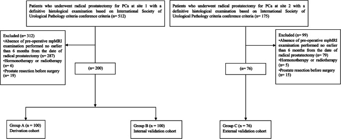
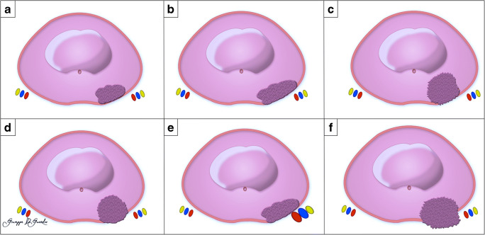
We retrospectively reviewed all consecutive patients admitted to two institutions for radical prostatectomy for PCa with available records of mpMRI performed between January 2015 and December 2020. Data from one institution was used for investigating diagnostic performance of each mEPE feature using radical prostatectomy specimens as benchmark. The results were implemented in a mEPE-score as follows: no mEPE features: 1; capsular abutment: 2; irregular or spiculated margin: 3; bulging prostatic contour, or asymmetry of the neurovascular bundles, or tumor-capsule interface > 1.0 cm: 4; ≥ 2 of the previous three parameters or measurable extraprostatic disease: 5. The performance of mEPE features was evaluated using the five diagnostic parameters and ROC curve analysis.

RESULTS

Two-hundred patients were enrolled at site 1 and 76 at site 2. mEPE features had poor sensitivities ranging from 0.08 (0.00-0.15) to 0.71 (0.59-0.83), whereas specificity ranged from 0.68 (0.58-0.79) to 1.00. mEPE-score showed excellent discriminating ability (AUC > 0.8) and sensitivity = 0.82 and specificity = 0.77 with a threshold of 3. mEPE-score had AUC comparable to ESUR-score (p = 0.59 internal validation; p = 0.82 external validation), higher than or comparable to mEPE-grade (p = 0.04 internal validation; p = 0.58 external validation), and higher than early-and-late-EPE (p < 0.0001 internal and external validation). There were no significant differences between readers having different expertise with EPE-score (p = 0.32) or mEPE-grade (p = 0.45), but there were significant differences for ESUR-score (p = 0.02) and early-versus-late-EPE (p = 0.03).

CONCLUSIONS

The individual mEPE features have low sensitivity and high specificity. The use of mEPE-score allows for consistent and reliable assessment for pathologic EPE.

KEY POINTS

• Individual PI-RADS v2.1 mpMRI features had poor sensitivities ranging from 0.08 (0.00-0.15) to 0.71 (0.59-0.83), whereas Sp ranged from 0.68 (0.58-0.79) to 1.00. • mEPE-score is an all-inclusive score for the assessment of pEPE with excellent discriminating ability (i.e., AUC > 0.8) and Se = 0.82, Sp = 0.77, PPV = 0.74, and NPV = 0.84 with a threshold of 3. • The diagnostic performance of the expert reader and beginner reader with pEPE-score was comparable (p = 0.32).

摘要

目的

研究 PI-RADS v2.1 多参数磁共振成像(mpMRI)特征在预测前列腺癌(PCa)前列腺外延伸(mEPE)中的诊断准确性,并开发和验证综合的 mpMRI 衍生评分(mEPE-score)。

方法

我们回顾性分析了 2015 年 1 月至 2020 年 12 月期间在两个机构因 PCa 接受根治性前列腺切除术且有 mpMRI 记录的所有连续患者。一家机构的数据用于使用根治性前列腺切除术标本作为基准来研究每个 mEPE 特征的诊断性能。结果在 mEPE-score 中实施如下:无 mEPE 特征:1;包膜侵犯:2;不规则或刺状边缘:3;前列腺轮廓隆起,或神经血管束不对称,或肿瘤包膜界面>1.0cm:4;前三个参数中的≥2 个或可测量的前列腺外疾病:5。使用五个诊断参数和 ROC 曲线分析评估 mEPE 特征的性能。

结果

site1 纳入了 200 例患者,site2 纳入了 76 例患者。mEPE 特征的敏感性范围从 0.08(0.00-0.15)到 0.71(0.59-0.83),特异性范围从 0.68(0.58-0.79)到 1.00。mEPE-score 具有出色的鉴别能力(AUC>0.8)和敏感性=0.82,特异性=0.77,阈值为 3。mEPE-score 的 AUC 与 ESUR-score 相当(p=0.59 内部验证;p=0.82 外部验证),高于或与 mEPE-grade 相当(p=0.04 内部验证;p=0.58 外部验证),高于早期和晚期 EPE(p<0.0001 内部和外部验证)。具有不同 EPE-score 经验的读者之间(p=0.32)或 mEPE-grade(p=0.45)没有显著差异,但 ESUR-score(p=0.02)和早期与晚期 EPE(p=0.03)之间存在显著差异。

结论

个体 mEPE 特征的敏感性较低,特异性较高。使用 mEPE-score 可以对病理 EPE 进行一致且可靠的评估。

关键点

  1. 个体 PI-RADS v2.1 mpMRI 特征的敏感性范围从 0.08(0.00-0.15)到 0.71(0.59-0.83),特异性范围从 0.68(0.58-0.79)到 1.00。

  2. mEPE-score 是一种全面的评估 pEPE 的评分,具有出色的鉴别能力(即 AUC>0.8)和敏感性=0.82,特异性=0.77,PPV=0.74,NPV=0.84,阈值为 3。

  3. 专家读者和初学者读者使用 pEPE-score 的诊断性能相当(p=0.32)。

https://cdn.ncbi.nlm.nih.gov/pmc/blobs/7c72/9213375/d28c15a03eb5/330_2022_8595_Fig6_HTML.jpg
https://cdn.ncbi.nlm.nih.gov/pmc/blobs/7c72/9213375/d6508a5bcbb3/330_2022_8595_Fig1_HTML.jpg
https://cdn.ncbi.nlm.nih.gov/pmc/blobs/7c72/9213375/5702d937f522/330_2022_8595_Fig2_HTML.jpg
https://cdn.ncbi.nlm.nih.gov/pmc/blobs/7c72/9213375/9cc9d10aa17f/330_2022_8595_Fig3_HTML.jpg
https://cdn.ncbi.nlm.nih.gov/pmc/blobs/7c72/9213375/003cb8cc7144/330_2022_8595_Fig4_HTML.jpg
https://cdn.ncbi.nlm.nih.gov/pmc/blobs/7c72/9213375/054f7ad1eb87/330_2022_8595_Fig5_HTML.jpg
https://cdn.ncbi.nlm.nih.gov/pmc/blobs/7c72/9213375/d28c15a03eb5/330_2022_8595_Fig6_HTML.jpg

相似文献

1
mEPE-score: a comprehensive grading system for predicting pathologic extraprostatic extension of prostate cancer at multiparametric magnetic resonance imaging.mEPE 评分:一种用于预测前列腺癌多参数磁共振成像下病理性前列腺外延伸的综合分级系统。
Eur Radiol. 2022 Jul;32(7):4942-4953. doi: 10.1007/s00330-022-08595-9. Epub 2022 Mar 15.
2
[Diagnostic value of multiparametric MRI-based models in the assessment of extra-prostatic extension of prostate cancer].[基于多参数磁共振成像的模型在评估前列腺癌前列腺外侵犯中的诊断价值]
Zhonghua Yi Xue Za Zhi. 2023 May 23;103(19):1439-1445. doi: 10.3760/cma.j.cn112137-20221215-02656.
3
Combined Clinical Parameters and Multiparametric Magnetic Resonance Imaging for the Prediction of Extraprostatic Disease-A Risk Model for Patient-tailored Risk Stratification When Planning Radical Prostatectomy.联合临床参数和多参数磁共振成像预测前列腺外疾病-用于计划根治性前列腺切除术时患者个体化风险分层的风险模型。
Eur Urol Focus. 2020 Nov 15;6(6):1205-1212. doi: 10.1016/j.euf.2018.11.004. Epub 2018 Nov 23.
4
Low PI-RADS assessment category excludes extraprostatic extension (≥pT3a) of prostate cancer: a histology-validated study including 301 operated patients.低 PI-RADS 评估类别排除前列腺癌的前列腺外延伸(≥pT3a):一项包括 301 例手术患者的组织学验证研究。
Eur Radiol. 2019 Oct;29(10):5478-5487. doi: 10.1007/s00330-019-06092-0. Epub 2019 Mar 18.
5
MR-based simplified extraprostatic extension evaluation: comparison of performances of different predictive models.基于磁共振成像的简化前列腺外扩展评估:不同预测模型的性能比较
Eur Radiol. 2023 Apr;33(4):2975-2984. doi: 10.1007/s00330-022-09240-1. Epub 2022 Dec 13.
6
Which one is better for predicting extraprostatic extension on multiparametric MRI: ESUR score, Likert scale, tumor contact length, or EPE grade?在多参数 MRI 上,哪种方法更能预测前列腺外延伸:ESUR 评分、Likert 量表、肿瘤接触长度还是 EPE 分级?
Eur J Radiol. 2022 Apr;149:110228. doi: 10.1016/j.ejrad.2022.110228. Epub 2022 Feb 23.
7
Extraprostatic Tumor Extension: Comparison of Preoperative Multiparametric MRI Criteria and Histopathologic Correlation after Radical Prostatectomy.前列腺外肿瘤侵犯:根治性前列腺切除术前多参数 MRI 标准与术后组织病理学相关性比较。
Radiology. 2020 Jul;296(1):87-95. doi: 10.1148/radiol.2020192133. Epub 2020 May 5.
8
How Can We Identify Extraprostatic Extension (EPE) Before Surgery? The Use of a Preoperative Prostate MRI EPE Scoring System to Assess Postprostatectomy Locally Advanced Prostate Cancer.我们如何在手术前确定前列腺外扩展(EPE)?术前前列腺 MRI EPE 评分系统在评估前列腺癌术后局部进展中的应用。
J Endourol. 2024 May;38(5):499-504. doi: 10.1089/end.2023.0572. Epub 2024 Mar 14.
9
A Grading System for the Assessment of Risk of Extraprostatic Extension of Prostate Cancer at Multiparametric MRI.前列腺癌多参数 MRI 外生评估风险的分级系统。
Radiology. 2019 Mar;290(3):709-719. doi: 10.1148/radiol.2018181278. Epub 2019 Jan 22.
10
Prostate Cancer: The European Society of Urogenital Radiology Prostate Imaging Reporting and Data System Criteria for Predicting Extraprostatic Extension by Using 3-T Multiparametric MR Imaging.前列腺癌:欧洲泌尿生殖放射学会前列腺成像报告和数据系统标准,用于预测使用 3T 多参数磁共振成像的前列腺外扩展。
Radiology. 2015 Aug;276(2):479-89. doi: 10.1148/radiol.15141412. Epub 2015 Apr 3.

引用本文的文献

1
10 mm (PI-RADS v2.1) versus 15 mm (PI-RADS v1.0) tumor capsule contact length in predicting extracapsular extension in prostate cancer: Meta-analysis and systematic review.10毫米(PI-RADS v2.1)与15毫米(PI-RADS v1.0)肿瘤包膜接触长度对预测前列腺癌包膜外侵犯的比较:荟萃分析与系统评价
Abdom Radiol (NY). 2025 Jun 5. doi: 10.1007/s00261-025-04998-6.
2
Lesion-based grading system using clinicopathological and MRI features for predicting positive surgical margins in prostate cancer.使用临床病理和MRI特征的基于病变的分级系统预测前列腺癌手术切缘阳性情况
Abdom Radiol (NY). 2025 Jan 28. doi: 10.1007/s00261-025-04808-z.
3

本文引用的文献

1
Prostate cancer local staging using biparametric MRI: assessment and comparison with multiparametric MRI.使用双参数 MRI 进行前列腺癌局部分期:评估及与多参数 MRI 的比较。
Eur J Radiol. 2020 Nov;132:109350. doi: 10.1016/j.ejrad.2020.109350. Epub 2020 Oct 15.
2
Prostate Imaging Quality (PI-QUAL): A New Quality Control Scoring System for Multiparametric Magnetic Resonance Imaging of the Prostate from the PRECISION trial.前列腺成像质量(PI-QUAL):来自 PRECISION 试验的前列腺多参数磁共振成像的新质量控制评分系统。
Eur Urol Oncol. 2020 Oct;3(5):615-619. doi: 10.1016/j.euo.2020.06.007. Epub 2020 Jul 6.
3
Prostate cancer: ESMO Clinical Practice Guidelines for diagnosis, treatment and follow-up.
Prognostic significance of the mEPE score in intermediate-risk prostate cancer patients undergoing ultrahypofractionated robotic SBRT.
mEPE评分在接受超分割机器人立体定向放疗的中危前列腺癌患者中的预后意义。
Strahlenther Onkol. 2025 Jan 14. doi: 10.1007/s00066-024-02355-y.
4
A preoperative scoring system for predicting the extraprostatic extension of prostate cancer following radical prostatectomy using magnetic resonance imaging and clinical factors.一种使用磁共振成像和临床因素预测前列腺癌根治性前列腺切除术后前列腺外侵犯的术前评分系统。
Abdom Radiol (NY). 2024 Aug;49(8):2683-2692. doi: 10.1007/s00261-024-04345-1. Epub 2024 May 16.
5
A pictorial essay of PI-RADS pearls and pitfalls: toward less ambiguity and better practice.PI-RADS要点与陷阱的图文并茂文章:力求减少模糊性并优化实践。
Abdom Radiol (NY). 2024 Sep;49(9):3190-3205. doi: 10.1007/s00261-024-04273-0. Epub 2024 May 5.
6
Extra-prostatic extension grading system: correlation with MRI features and integration of capsular enhancement sign for "enhanced" detection of T3a lesions.前列腺外延伸分级系统:与 MRI 特征的相关性以及包膜增强征象的整合,用于“增强”检测 T3a 病变。
Br J Radiol. 2024 May 7;97(1157):971-979. doi: 10.1093/bjr/tqae065.
7
Effect of preoperative PI-RADS assessment on pathological outcomes in patients who underwent radical prostatectomy.术前 PI-RADS 评估对接受根治性前列腺切除术患者病理结果的影响。
Cancer Imaging. 2023 Nov 26;23(1):113. doi: 10.1186/s40644-023-00619-x.
8
Radiomics for the identification of extraprostatic extension with prostate MRI: a systematic review and meta-analysis.基于 MRI 的前列腺癌外扩的影像组学分析:一项系统综述和荟萃分析。
Eur Radiol. 2024 Jun;34(6):3981-3991. doi: 10.1007/s00330-023-10427-3. Epub 2023 Nov 13.
9
Imaging features of the PI-RADS for predicting extraprostatic extension of prostate cancer: systematic review and meta-analysis.前列腺影像报告和数据系统(PI-RADS)预测前列腺癌前列腺外侵犯的影像特征:系统评价与荟萃分析
Insights Imaging. 2023 May 8;14(1):77. doi: 10.1186/s13244-023-01422-9.
10
Role of the Prostate Imaging Quality PI-QUAL Score for Prostate Magnetic Resonance Image Quality in Pathological Upstaging After Radical Prostatectomy: A Multicentre European Study.前列腺成像质量PI-QUAL评分在前列腺癌根治术后病理分期升级中对前列腺磁共振图像质量的作用:一项多中心欧洲研究
Eur Urol Open Sci. 2022 Dec 15;47:94-101. doi: 10.1016/j.euros.2022.11.013. eCollection 2023 Jan.
前列腺癌:欧洲肿瘤内科学会临床实践指南之诊断、治疗及随访
Ann Oncol. 2020 Sep;31(9):1119-1134. doi: 10.1016/j.annonc.2020.06.011. Epub 2020 Jun 25.
4
ESUR/ESUI consensus statements on multi-parametric MRI for the detection of clinically significant prostate cancer: quality requirements for image acquisition, interpretation and radiologists' training.ESUR/ESUI 共识声明:多参数 MRI 检测临床显著前列腺癌:图像采集、解读和放射科医生培训的质量要求。
Eur Radiol. 2020 Oct;30(10):5404-5416. doi: 10.1007/s00330-020-06929-z. Epub 2020 May 19.
5
The Diagnostic Performance of the Length of Tumor Capsular Contact on MRI for Detecting Prostate Cancer Extraprostatic Extension: A Systematic Review and Meta-Analysis.MRI 上肿瘤包膜接触长度诊断前列腺癌前列腺外侵犯的效能:系统评价和荟萃分析。
Korean J Radiol. 2020 Jun;21(6):684-694. doi: 10.3348/kjr.2019.0842.
6
T-staging of prostate cancer: Identification of useful signs to standardize detection of posterolateral extraprostatic extension on prostate MRI.前列腺癌 T 分期:识别有用的征象,以标准化前列腺 MRI 检测后外侧前列腺外延伸。
Clin Imaging. 2020 Jan;59(1):1-7. doi: 10.1016/j.clinimag.2019.08.007. Epub 2019 Sep 13.
7
Machine learning applications in prostate cancer magnetic resonance imaging.机器学习在前列腺癌磁共振成像中的应用。
Eur Radiol Exp. 2019 Aug 7;3(1):35. doi: 10.1186/s41747-019-0109-2.
8
Head-to-head comparison between multiparametric MRI, the partin tables, memorial sloan kettering cancer center nomogram, and CAPRA score in predicting extraprostatic cancer in patients undergoing radical prostatectomy.多参数 MRI、Partin 表、纪念斯隆-凯特琳癌症中心列线图和 CAPRA 评分在预测接受根治性前列腺切除术患者前列腺外癌中的头对头比较。
J Magn Reson Imaging. 2019 Nov;50(5):1604-1613. doi: 10.1002/jmri.26743. Epub 2019 Apr 7.
9
A Grading System for the Assessment of Risk of Extraprostatic Extension of Prostate Cancer at Multiparametric MRI.前列腺癌多参数 MRI 外生评估风险的分级系统。
Radiology. 2019 Mar;290(3):709-719. doi: 10.1148/radiol.2018181278. Epub 2019 Jan 22.
10
Combined Clinical Parameters and Multiparametric Magnetic Resonance Imaging for the Prediction of Extraprostatic Disease-A Risk Model for Patient-tailored Risk Stratification When Planning Radical Prostatectomy.联合临床参数和多参数磁共振成像预测前列腺外疾病-用于计划根治性前列腺切除术时患者个体化风险分层的风险模型。
Eur Urol Focus. 2020 Nov 15;6(6):1205-1212. doi: 10.1016/j.euf.2018.11.004. Epub 2018 Nov 23.