• 文献检索
  • 文档翻译
  • 深度研究
  • 学术资讯
  • Suppr Zotero 插件Zotero 插件
  • 邀请有礼
  • 套餐&价格
  • 历史记录
应用&插件
Suppr Zotero 插件Zotero 插件浏览器插件Mac 客户端Windows 客户端微信小程序
定价
高级版会员购买积分包购买API积分包
服务
文献检索文档翻译深度研究API 文档MCP 服务
关于我们
关于 Suppr公司介绍联系我们用户协议隐私条款
关注我们

Suppr 超能文献

核心技术专利:CN118964589B侵权必究
粤ICP备2023148730 号-1Suppr @ 2026

文献检索

告别复杂PubMed语法,用中文像聊天一样搜索,搜遍4000万医学文献。AI智能推荐,让科研检索更轻松。

立即免费搜索

文件翻译

保留排版,准确专业,支持PDF/Word/PPT等文件格式,支持 12+语言互译。

免费翻译文档

深度研究

AI帮你快速写综述,25分钟生成高质量综述,智能提取关键信息,辅助科研写作。

立即免费体验

眼肌 MRI 在重症肌无力和其他神经肌肉疾病中的应用。

Eye Muscle MRI in Myasthenia Gravis and Other Neuromuscular Disorders.

机构信息

Department of Radiology, CJ Gorter MRI Center, Leiden University Medical Center, Leiden, The Netherlands.

Department of Neurology, Leiden University Medical Center, Leiden, The Netherlands.

出版信息

J Neuromuscul Dis. 2023;10(5):869-883. doi: 10.3233/JND-230023.

DOI:10.3233/JND-230023
PMID:37182896
原文链接:https://pmc.ncbi.nlm.nih.gov/articles/PMC10578256/
Abstract

INTRODUCTION

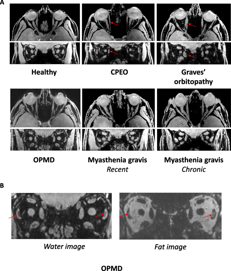
MRI of extra-ocular muscles (EOM) in patients with myasthenia gravis (MG) could aid in diagnosis and provide insights in therapy-resistant ophthalmoplegia. We used quantitative MRI to study the EOM in MG, healthy and disease controls, including Graves' ophthalmopathy (GO), oculopharyngeal muscular dystrophy (OPMD) and chronic progressive external ophthalmoplegia (CPEO).

METHODS

Twenty recently diagnosed MG (59±19yrs), nineteen chronic MG (51±16yrs), fourteen seronegative MG (57±9yrs) and sixteen healthy controls (54±13yrs) were included. Six CPEO (49±14yrs), OPMD (62±10yrs) and GO patients (44±12yrs) served as disease controls. We quantified muscle fat fraction (FF), T2water and volume. Eye ductions and gaze deviations were assessed by synoptophore and Hess-charting.

RESULTS

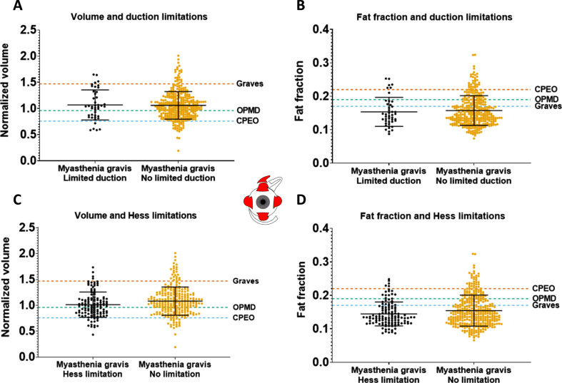
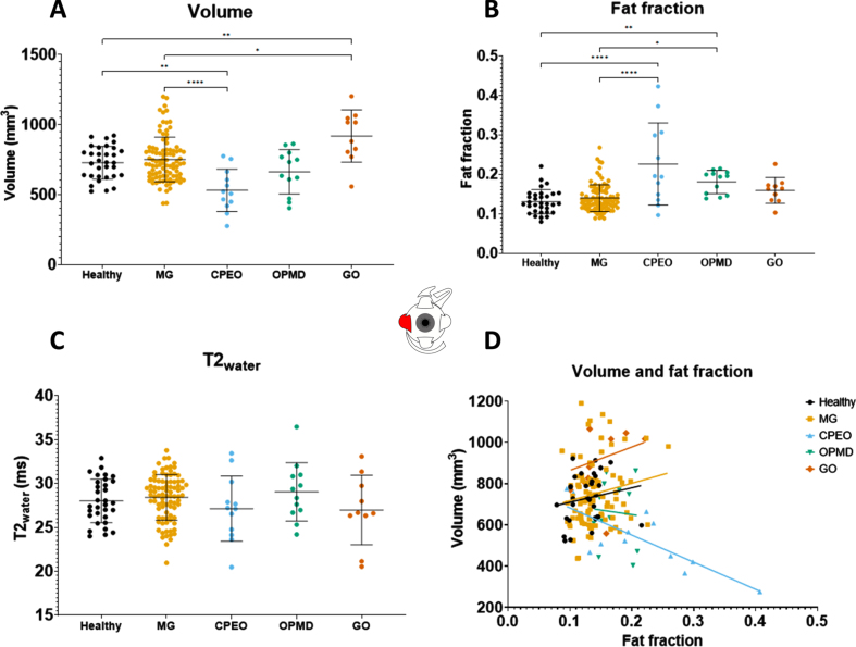
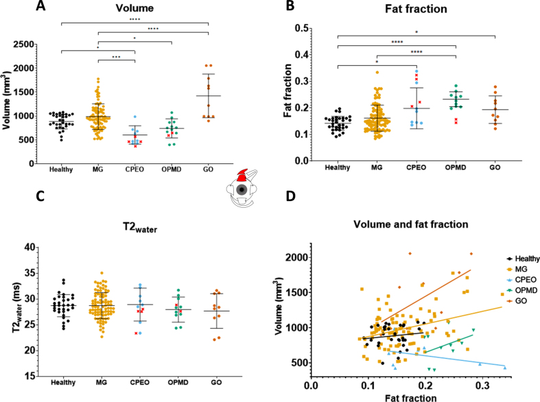
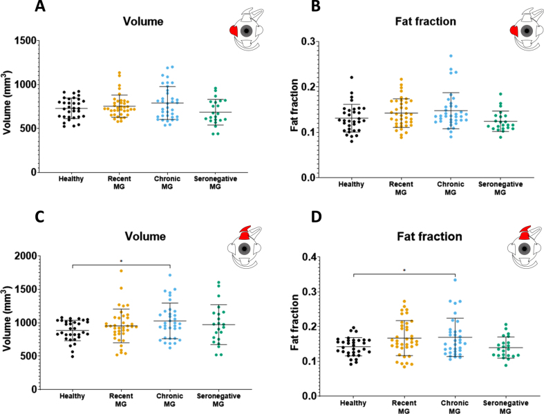
Chronic, but not recent onset, MG patients showed volume increases (e.g. superior rectus and levator palpebrae [SR+LPS] 985±155 mm3 compared to 884±269 mm3 for healthy controls, p < 0.05). As expected, in CPEO volume was decreased (e.g. SR+LPS 602±193 mm3, p < 0.0001), and in GO volume was increased (e.g. SR+LPS 1419±457 mm3, p < 0.0001). FF was increased in chronic MG (e.g. medial rectus increased 0.017, p < 0.05). In CPEO and OPMD the FF was more severely increased. The severity of ophthalmoplegia did not correlate with EOM volume in MG, but did in CPEO and OPMD. No differences in T2water were found.

INTERPRETATION

We observed small increases in EOM volume and FF in chronic MG compared to healthy controls. Surprisingly, we found no atrophy in MG, even in patients with long-term ophthalmoplegia. This implies that even long-term ophthalmoplegia in MG does not lead to secondary structural myopathic changes precluding functional recovery.

摘要

介绍

对重症肌无力(MG)患者的眼外肌(EOM)进行 MRI 检查有助于诊断,并为治疗抵抗性眼肌麻痹提供深入了解。我们使用定量 MRI 研究了 MG、健康对照者和疾病对照组(包括格雷夫斯眼病(GO)、眼咽肌营养不良(OPMD)和慢性进行性眼外肌麻痹(CPEO))的 EOM。

方法

共纳入 20 例新近诊断的 MG(59±19 岁)、19 例慢性 MG(51±16 岁)、14 例血清阴性 MG(57±9 岁)和 16 例健康对照者(54±13 岁)。6 例 CPEO(49±14 岁)、6 例 OPMD(62±10 岁)和 6 例 GO 患者(44±12 岁)作为疾病对照组。我们定量了肌肉脂肪分数(FF)、T2 水和体积。使用同视机和 Hess 图表评估眼球运动和凝视偏差。

结果

慢性但非近期发病的 MG 患者显示出体积增加(例如,上直肌和提上睑肌[SR+LPS]为 985±155mm3,而健康对照组为 884±269mm3,p<0.05)。如预期的那样,CPEO 患者的体积减少(例如,SR+LPS 为 602±193mm3,p<0.0001),GO 患者的体积增加(例如,SR+LPS 为 1419±457mm3,p<0.0001)。慢性 MG 患者的 FF 增加(例如,内侧直肌增加 0.017,p<0.05)。CPEO 和 OPMD 患者的 FF 增加更为严重。MG 患者的眼肌麻痹严重程度与 EOM 体积无关,但在 CPEO 和 OPMD 患者中有关。未发现 T2 水的差异。

解释

与健康对照组相比,我们观察到慢性 MG 患者的 EOM 体积和 FF 略有增加。令人惊讶的是,即使在患有长期眼肌麻痹的患者中,我们也未发现 MG 有萎缩。这意味着即使是 MG 的长期眼肌麻痹也不会导致继发性结构性肌病改变,从而妨碍功能恢复。

https://cdn.ncbi.nlm.nih.gov/pmc/blobs/33e1/10578256/c6a42360581a/jnd-10-jnd230023-g005.jpg
https://cdn.ncbi.nlm.nih.gov/pmc/blobs/33e1/10578256/8f4a707b96d6/jnd-10-jnd230023-g001.jpg
https://cdn.ncbi.nlm.nih.gov/pmc/blobs/33e1/10578256/2a02dc5f9fe9/jnd-10-jnd230023-g002.jpg
https://cdn.ncbi.nlm.nih.gov/pmc/blobs/33e1/10578256/d79cae85952e/jnd-10-jnd230023-g003.jpg
https://cdn.ncbi.nlm.nih.gov/pmc/blobs/33e1/10578256/136f8bbca9c9/jnd-10-jnd230023-g004.jpg
https://cdn.ncbi.nlm.nih.gov/pmc/blobs/33e1/10578256/c6a42360581a/jnd-10-jnd230023-g005.jpg

相似文献

1
Eye Muscle MRI in Myasthenia Gravis and Other Neuromuscular Disorders.眼肌 MRI 在重症肌无力和其他神经肌肉疾病中的应用。
J Neuromuscul Dis. 2023;10(5):869-883. doi: 10.3233/JND-230023.
2
The feasibility of quantitative MRI of extra-ocular muscles in myasthenia gravis and Graves' orbitopathy.定量 MRI 在外眼肌型重症肌无力和格雷夫斯眼病中的可行性研究。
NMR Biomed. 2021 Jan;34(1):e4407. doi: 10.1002/nbm.4407. Epub 2020 Sep 7.
3
Clinical and imaging clues to the diagnosis and follow-up of ptosis and ophthalmoparesis.眼睑下垂和眼肌瘫痪的诊断和随访的临床及影像学线索。
J Cachexia Sarcopenia Muscle. 2022 Dec;13(6):2820-2834. doi: 10.1002/jcsm.13089. Epub 2022 Sep 29.
4
Orbital magnetic resonance imaging of extraocular muscles in chronic progressive external ophthalmoplegia: specific diagnostic findings.慢性进行性眼外肌麻痹患者眼外肌的眼眶磁共振成像:特异性诊断结果
J AAPOS. 2006 Oct;10(5):414-8. doi: 10.1016/j.jaapos.2006.04.012.
5
Extra-ocular muscle MRI in genetically-defined mitochondrial disease.基因明确的线粒体疾病的眼外肌磁共振成像
Eur Radiol. 2016 Jan;26(1):130-7. doi: 10.1007/s00330-015-3801-5. Epub 2015 May 21.
6
Diagnosing myasthenia gravis using orthoptic measurements: assessing extraocular muscle fatiguability.使用眼肌测量法诊断重症肌无力:评估眼外肌疲劳性
J Neurol Neurosurg Psychiatry. 2023 Feb;94(2):151. doi: 10.1136/jnnp-2022-329859. Epub 2022 Oct 19.
7
MR of extraocular muscles in chronic progressive external ophthalmoplegia.慢性进行性眼外肌麻痹的眼外肌磁共振成像
AJNR Am J Neuroradiol. 1998 Jan;19(1):95-9.
8
Extraocular Muscle Findings in Myasthenia Gravis Associated Treatment-Resistant Ophthalmoplegia.重症肌无力相关治疗抵抗性眼肌麻痹的眼外肌表现
J Neuroophthalmol. 2017 Dec;37(4):414-417. doi: 10.1097/WNO.0000000000000534.
9
MRI evidence of extraocular muscle atrophy and fatty replacement in myasthenia gravis.MRI 证据显示重症肌无力患者眼外肌萎缩和脂肪替代。
Neuroradiology. 2021 Sep;63(9):1531-1538. doi: 10.1007/s00234-021-02753-4. Epub 2021 Jul 7.
10
Eyelid ptosis (Blepharoptosis) for the primary care practitioner.初级保健医生须知的眼睑下垂(上睑下垂)
Dis Mon. 2020 Oct;66(10):101040. doi: 10.1016/j.disamonth.2020.101040. Epub 2020 Jun 27.

引用本文的文献

1
Novel Device for Intraoperative Quantitative Measurements of Extraocular Muscle Tensile Strength.用于术中定量测量眼外肌拉伸强度的新型装置。
Biosensors (Basel). 2025 May 30;15(6):347. doi: 10.3390/bios15060347.
2
Diaphragm ultrasound and neostigmine test as complementary tools in the diagnostic assessment of myasthenia gravis: insights from a clinical case.膈肌超声和新斯的明试验作为重症肌无力诊断评估的辅助工具:来自临床病例的见解
Neurol Sci. 2025 Jun 13. doi: 10.1007/s10072-025-08264-x.
3
Rituximab for a rare pediatric case of concurrent thyroid eye disease and myasthenia gravis.

本文引用的文献

1
Advocating Patient-Centred Research in Ocular Myasthenia Gravis (OMG): A Call for an OMG Research Consortium.倡导以患者为中心的重症肌无力性眼病(OMG)研究:呼吁成立OMG研究联盟。
Front Ophthalmol (Lausanne). 2022 Jul 12;2:912805. doi: 10.3389/fopht.2022.912805. eCollection 2022.
2
Diagnosing myasthenia gravis using orthoptic measurements: assessing extraocular muscle fatiguability.使用眼肌测量法诊断重症肌无力:评估眼外肌疲劳性
J Neurol Neurosurg Psychiatry. 2023 Feb;94(2):151. doi: 10.1136/jnnp-2022-329859. Epub 2022 Oct 19.
3
Extraocular Muscle Atrophy in Myasthenia Gravis.
利妥昔单抗治疗罕见的小儿甲状腺眼病合并重症肌无力病例
Ther Adv Endocrinol Metab. 2025 May 28;16:20420188251340137. doi: 10.1177/20420188251340137. eCollection 2025.
4
A Focus on Myasthenic Ptosis: The Interface of Medical and Surgical Treatment.聚焦重症肌无力性上睑下垂:药物治疗与手术治疗的衔接
World J Plast Surg. 2024;13(3):23-32. doi: 10.61186/wjps.13.3.23.
5
Myasthenia gravis: the future is here.重症肌无力:未来已来。
J Clin Invest. 2024 Jun 17;134(12):e179742. doi: 10.1172/JCI179742.
重症肌无力中的眼外肌萎缩
Ann Indian Acad Neurol. 2022 May-Jun;25(3):483-484. doi: 10.4103/aian.AIAN_1039_21. Epub 2022 Apr 7.
4
Epidemiology, diagnostics, and biomarkers of autoimmune neuromuscular junction disorders.自身免疫性神经肌肉接头疾病的流行病学、诊断和生物标志物。
Lancet Neurol. 2022 Feb;21(2):176-188. doi: 10.1016/S1474-4422(21)00297-0.
5
Eye-specific quantitative dynamic contrast-enhanced MRI analysis for patients with intraocular masses.眼部特异性定量动态对比增强 MRI 分析在眼内肿块患者中的应用。
MAGMA. 2022 Apr;35(2):311-323. doi: 10.1007/s10334-021-00961-w. Epub 2021 Oct 13.
6
MRI evidence of extraocular muscle atrophy and fatty replacement in myasthenia gravis.MRI 证据显示重症肌无力患者眼外肌萎缩和脂肪替代。
Neuroradiology. 2021 Sep;63(9):1531-1538. doi: 10.1007/s00234-021-02753-4. Epub 2021 Jul 7.
7
Challenges in diagnosing coexisting ocular myasthenia gravis and thyroid eye disease.诊断共存性眼肌型重症肌无力和甲状腺眼病的挑战。
Muscle Nerve. 2021 May;63(5):631-639. doi: 10.1002/mus.27118. Epub 2020 Nov 27.
8
The feasibility of quantitative MRI of extra-ocular muscles in myasthenia gravis and Graves' orbitopathy.定量 MRI 在外眼肌型重症肌无力和格雷夫斯眼病中的可行性研究。
NMR Biomed. 2021 Jan;34(1):e4407. doi: 10.1002/nbm.4407. Epub 2020 Sep 7.
9
T relaxation-time mapping in healthy and diseased skeletal muscle using extended phase graph algorithms.利用扩展相图算法对健康和患病骨骼肌中的 T 弛豫时间进行映射。
Magn Reson Med. 2020 Nov;84(5):2656-2670. doi: 10.1002/mrm.28290. Epub 2020 Apr 19.
10
The Muscle Is Not a Passive Target in Myasthenia Gravis.在重症肌无力中,肌肉并非被动靶点。
Front Neurol. 2019 Dec 19;10:1343. doi: 10.3389/fneur.2019.01343. eCollection 2019.