• 文献检索
  • 文档翻译
  • 深度研究
  • 学术资讯
  • Suppr Zotero 插件Zotero 插件
  • 邀请有礼
  • 套餐&价格
  • 历史记录
应用&插件
Suppr Zotero 插件Zotero 插件浏览器插件Mac 客户端Windows 客户端微信小程序
定价
高级版会员购买积分包购买API积分包
服务
文献检索文档翻译深度研究API 文档MCP 服务
关于我们
关于 Suppr公司介绍联系我们用户协议隐私条款
关注我们

Suppr 超能文献

核心技术专利:CN118964589B侵权必究
粤ICP备2023148730 号-1Suppr @ 2026

文献检索

告别复杂PubMed语法,用中文像聊天一样搜索,搜遍4000万医学文献。AI智能推荐,让科研检索更轻松。

立即免费搜索

文件翻译

保留排版,准确专业,支持PDF/Word/PPT等文件格式,支持 12+语言互译。

免费翻译文档

深度研究

AI帮你快速写综述,25分钟生成高质量综述,智能提取关键信息,辅助科研写作。

立即免费体验

ErbB 信号转导是伴有纤维成分的血管病变的潜在治疗靶点。

ErbB signaling is a potential therapeutic target for vascular lesions with fibrous component.

机构信息

A.I. Virtanen Institute for Molecular Sciences, University of Eastern Finland, Kuopio, Finland.

Department of Plastic Surgery, Helsinki University Hospital and University of Helsinki, Helsinki, Finland.

出版信息

Elife. 2023 May 18;12:e82543. doi: 10.7554/eLife.82543.

DOI:10.7554/eLife.82543
PMID:37199488
原文链接:https://pmc.ncbi.nlm.nih.gov/articles/PMC10260011/
Abstract

BACKGROUND

Sporadic venous malformation (VM) and angiomatosis of soft tissue (AST) are benign, congenital vascular anomalies affecting venous vasculature. Depending on the size and location of the lesion, symptoms vary from motility disturbances to pain and disfigurement. Due to the high recurrence of the lesions, more effective therapies are needed.

METHODS

As targeting stromal cells has been an emerging concept in anti-angiogenic therapies, here, by using VM/AST patient samples, RNA-sequencing, cell culture techniques, and a xenograft mouse model, we investigated the crosstalk of endothelial cells (EC) and fibroblasts and its effect on vascular lesion growth.

RESULTS

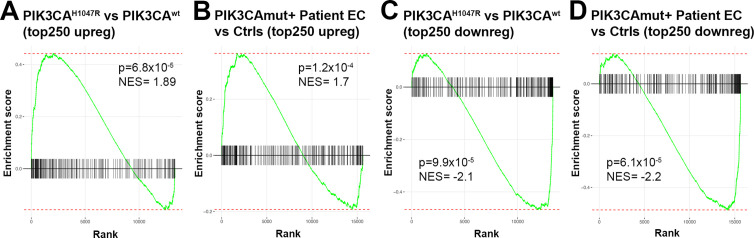
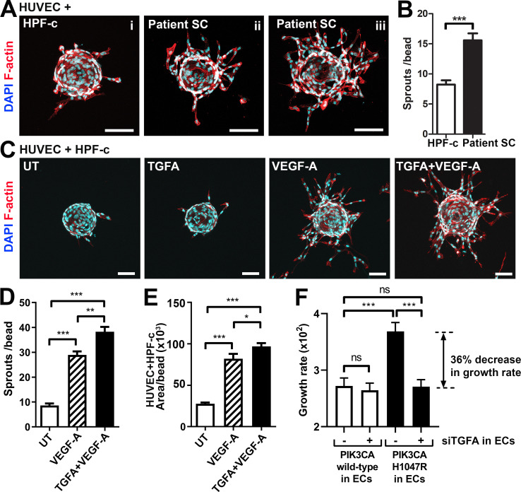
We report, for the first time, the expression and secretion of transforming growth factor A (TGFA) in ECs or intervascular stromal cells in AST and VM lesions. TGFA induced secretion of vascular endothelial growth factor (VEGF-A) in paracrine fashion, and regulated EC proliferation. Oncogenic variant in p.H1047R, a common somatic mutation found in these lesions, increased TGFA expression, enrichment of hallmark hypoxia, and in a mouse xenograft model, lesion size, and vascularization. Treatment with afatinib, a pan-ErbB tyrosine-kinase inhibitor, decreased vascularization and lesion size in a mouse xenograft model with ECs expressing oncogenic p.H1047R variant and fibroblasts.

CONCLUSIONS

Based on the data, we suggest that targeting of both intervascular stromal cells and ECs is a potential treatment strategy for vascular lesions having a fibrous component.

FUNDING

Academy of Finland, Ella and Georg Ehnrooth foundation, the ERC grants, Sigrid Jusélius Foundation, Finnish Foundation for Cardiovascular Research, Jane and Aatos Erkko Foundation, GeneCellNano Flagship program, and Department of Musculoskeletal and Plastic Surgery, Helsinki University Hospital.

摘要

背景

散发性静脉畸形(VM)和软组织血管瘤病(AST)是良性的先天性血管异常,影响静脉血管系统。根据病变的大小和位置,症状从运动障碍到疼痛和畸形不等。由于病变的高复发率,需要更有效的治疗方法。

方法

由于靶向基质细胞已成为抗血管生成治疗的一个新兴概念,在这里,我们使用 VM/AST 患者样本、RNA 测序、细胞培养技术和异种移植小鼠模型,研究了内皮细胞(EC)和成纤维细胞之间的串扰及其对血管病变生长的影响。

结果

我们首次报道了转化生长因子 A(TGFA)在 AST 和 VM 病变中的 EC 或血管间基质细胞中的表达和分泌。TGFA 以旁分泌的方式诱导血管内皮生长因子(VEGF-A)的分泌,并调节 EC 的增殖。在这些病变中发现的常见体细胞突变 p.H1047R 的致癌变体增加了 TGFA 的表达、标志性缺氧的富集,并且在小鼠异种移植模型中增加了病变大小和血管化。泛-ErbB 酪氨酸激酶抑制剂 afatinib 的治疗降低了表达致癌 p.H1047R 变体的 EC 和成纤维细胞的小鼠异种移植模型中的血管化和病变大小。

结论

基于这些数据,我们建议针对具有纤维成分的血管病变的血管间基质细胞和 EC 的靶向治疗策略是一种潜在的治疗策略。

资助

芬兰科学院、Ella 和 Georg Ehnrooth 基金会、ERC 拨款、Sigrid Jusélius 基金会、芬兰心血管研究基金会、Jane 和 Aatos Erkko 基金会、GeneCellNano 旗舰计划以及赫尔辛基大学医院肌肉骨骼和整形外科。

https://cdn.ncbi.nlm.nih.gov/pmc/blobs/b527/10260011/8fd08cb64a0f/elife-82543-fig8.jpg
https://cdn.ncbi.nlm.nih.gov/pmc/blobs/b527/10260011/f756a4605d1b/elife-82543-fig1.jpg
https://cdn.ncbi.nlm.nih.gov/pmc/blobs/b527/10260011/dbe24af92b8d/elife-82543-fig1-figsupp1.jpg
https://cdn.ncbi.nlm.nih.gov/pmc/blobs/b527/10260011/23cbef906232/elife-82543-fig1-figsupp2.jpg
https://cdn.ncbi.nlm.nih.gov/pmc/blobs/b527/10260011/fb889065c1f8/elife-82543-fig2.jpg
https://cdn.ncbi.nlm.nih.gov/pmc/blobs/b527/10260011/37e01bcd9c35/elife-82543-fig2-figsupp1.jpg
https://cdn.ncbi.nlm.nih.gov/pmc/blobs/b527/10260011/ad4b1ec032b3/elife-82543-fig2-figsupp2.jpg
https://cdn.ncbi.nlm.nih.gov/pmc/blobs/b527/10260011/6c1901aeb078/elife-82543-fig2-figsupp3.jpg
https://cdn.ncbi.nlm.nih.gov/pmc/blobs/b527/10260011/f3bb79d00cb5/elife-82543-fig3.jpg
https://cdn.ncbi.nlm.nih.gov/pmc/blobs/b527/10260011/6fa27bff9d51/elife-82543-fig3-figsupp1.jpg
https://cdn.ncbi.nlm.nih.gov/pmc/blobs/b527/10260011/b78e224c3d70/elife-82543-fig3-figsupp2.jpg
https://cdn.ncbi.nlm.nih.gov/pmc/blobs/b527/10260011/440e64984f59/elife-82543-fig3-figsupp3.jpg
https://cdn.ncbi.nlm.nih.gov/pmc/blobs/b527/10260011/1edad19dfae2/elife-82543-fig4.jpg
https://cdn.ncbi.nlm.nih.gov/pmc/blobs/b527/10260011/c12b1d4da336/elife-82543-fig4-figsupp1.jpg
https://cdn.ncbi.nlm.nih.gov/pmc/blobs/b527/10260011/b1d0f55ed479/elife-82543-fig4-figsupp2.jpg
https://cdn.ncbi.nlm.nih.gov/pmc/blobs/b527/10260011/e62eb4898368/elife-82543-fig5.jpg
https://cdn.ncbi.nlm.nih.gov/pmc/blobs/b527/10260011/803d85c22bdc/elife-82543-fig5-figsupp1.jpg
https://cdn.ncbi.nlm.nih.gov/pmc/blobs/b527/10260011/a4d6e9fc5d41/elife-82543-fig6.jpg
https://cdn.ncbi.nlm.nih.gov/pmc/blobs/b527/10260011/65cdc6fca53e/elife-82543-fig7.jpg
https://cdn.ncbi.nlm.nih.gov/pmc/blobs/b527/10260011/8fd08cb64a0f/elife-82543-fig8.jpg

相似文献

1
ErbB signaling is a potential therapeutic target for vascular lesions with fibrous component.ErbB 信号转导是伴有纤维成分的血管病变的潜在治疗靶点。
Elife. 2023 May 18;12:e82543. doi: 10.7554/eLife.82543.
2
A xenograft model for venous malformation.静脉畸形的异种移植模型。
Angiogenesis. 2018 Nov;21(4):725-735. doi: 10.1007/s10456-018-9624-7. Epub 2018 May 21.
3
Somatic PIK3CA mutations as a driver of sporadic venous malformations.体细胞PIK3CA突变作为散发性静脉畸形的驱动因素。
Sci Transl Med. 2016 Mar 30;8(332):332ra42. doi: 10.1126/scitranslmed.aaf1164.
4
Constitutively active PIK3CA mutations are expressed by lymphatic and vascular endothelial cells in capillary lymphatic venous malformation.毛细血管淋巴管静脉畸形中淋巴管和血管内皮细胞表达组成性激活的 PIK3CA 突变。
Angiogenesis. 2020 Aug;23(3):425-442. doi: 10.1007/s10456-020-09722-0. Epub 2020 Apr 30.
5
PI3K/mTOR inhibition promotes the regression of experimental vascular malformations driven by PIK3CA-activating mutations.PI3K/mTOR 抑制促进由 PIK3CA 激活突变驱动的实验性血管畸形的消退。
Cell Death Dis. 2018 Jan 19;9(2):45. doi: 10.1038/s41419-017-0064-x.
6
A Xenograft Model for Venous Malformation.静脉畸形的异种移植模型。
Methods Mol Biol. 2021;2206:179-192. doi: 10.1007/978-1-0716-0916-3_13.
7
Ponatinib Combined With Rapamycin Causes Regression of Murine Venous Malformation.帕纳替尼联合雷帕霉素可导致鼠静脉畸形消退。
Arterioscler Thromb Vasc Biol. 2019 Mar;39(3):496-512. doi: 10.1161/ATVBAHA.118.312315.
8
Somatic activating mutations in Pik3ca cause sporadic venous malformations in mice and humans.磷脂酰肌醇-3激酶催化亚基α(PIK3CA)的体细胞激活突变可导致小鼠和人类出现散发性静脉畸形。
Sci Transl Med. 2016 Mar 30;8(332):332ra43. doi: 10.1126/scitranslmed.aad9982.
9
A Patient-Derived Xenograft Model for Venous Malformation.一种用于静脉畸形的患者来源异种移植模型。
J Vis Exp. 2020 Jun 15(160). doi: 10.3791/61501.
10
Somatic Activating PIK3CA Mutations Cause Venous Malformation.体细胞激活型PIK3CA突变导致静脉畸形。
Am J Hum Genet. 2015 Dec 3;97(6):914-21. doi: 10.1016/j.ajhg.2015.11.011.

引用本文的文献

1
Endothelial p.R183Q mutation confers hemoporfin-mediated photodynamic therapy resistance and drives pathological angiogenesis via the angiopoietin-2/TIE2/PI3K/AKT pathway.内皮细胞p.R183Q突变赋予血卟啉介导的光动力治疗抗性,并通过血管生成素-2/TIE2/PI3K/AKT途径驱动病理性血管生成。
Front Cell Dev Biol. 2025 Aug 18;13:1622961. doi: 10.3389/fcell.2025.1622961. eCollection 2025.
2
Embryological cellular origins and hypoxia-mediated mechanisms in PIK3CA-driven refractory vascular malformations.PIK3CA驱动的难治性血管畸形的胚胎细胞起源及缺氧介导机制
EMBO Mol Med. 2025 Apr 16. doi: 10.1038/s44321-025-00235-1.
3

本文引用的文献

1
SproutAngio: an open-source bioimage informatics tool for quantitative analysis of sprouting angiogenesis and lumen space.SproutAngio:一种开源的生物影像信息学工具,用于对发芽血管生成和管腔空间进行定量分析。
Sci Rep. 2023 May 4;13(1):7279. doi: 10.1038/s41598-023-33090-6.
2
Gene Set Knowledge Discovery with Enrichr.基因集知识发现与 Enrichr
Curr Protoc. 2021 Mar;1(3):e90. doi: 10.1002/cpz1.90.
3
BMP6/TAZ-Hippo signaling modulates angiogenesis and endothelial cell response to VEGF.BMP6/TAZ-Hippo 信号通路调节血管生成和内皮细胞对 VEGF 的反应。
Lymphatic malformations: mechanistic insights and evolving therapeutic frontiers.
淋巴管畸形:发病机制的深入理解和不断发展的治疗前沿。
J Clin Invest. 2024 Mar 15;134(6):e172844. doi: 10.1172/JCI172844.
Angiogenesis. 2021 Feb;24(1):129-144. doi: 10.1007/s10456-020-09748-4. Epub 2020 Oct 6.
4
The nf-core framework for community-curated bioinformatics pipelines.用于社区策划生物信息学流程的nf-core框架。
Nat Biotechnol. 2020 Mar;38(3):276-278. doi: 10.1038/s41587-020-0439-x.
5
Soft tissue angiomatosis: another PIK3CA-related disorder.软组织血管畸形:另一种与 PIK3CA 相关的疾病。
Histopathology. 2020 Mar;76(4):540-549. doi: 10.1111/his.14021. Epub 2020 Jan 24.
6
A xenograft model for venous malformation.静脉畸形的异种移植模型。
Angiogenesis. 2018 Nov;21(4):725-735. doi: 10.1007/s10456-018-9624-7. Epub 2018 May 21.
7
Treatment of Venous Malformations: The Data, Where We Are, and How It Is Done.静脉畸形的治疗:数据、现状及治疗方法
Tech Vasc Interv Radiol. 2018 Jun;21(2):45-54. doi: 10.1053/j.tvir.2018.03.001. Epub 2018 Mar 8.
8
Somatic Activating KRAS Mutations in Arteriovenous Malformations of the Brain.脑部动静脉畸形中的体细胞激活型KRAS突变
N Engl J Med. 2018 Apr 19;378(16):1561-1562. doi: 10.1056/NEJMc1802190.
9
Systematic Functional Annotation of Somatic Mutations in Cancer.癌症体细胞突变的系统功能注释。
Cancer Cell. 2018 Mar 12;33(3):450-462.e10. doi: 10.1016/j.ccell.2018.01.021.
10
Hypoxia-Preconditioned Human Umbilical Vein Endothelial Cells Protect Against Neurovascular Damage After Hypoxic Ischemia in Neonatal Brain.缺氧预处理人脐静脉内皮细胞对新生儿脑缺氧缺血后神经血管损伤的保护作用。
Mol Neurobiol. 2018 Oct;55(10):7743-7757. doi: 10.1007/s12035-018-0867-5. Epub 2018 Feb 19.