Lim Mark J, Yagnik Gargey, Henkel Corinna, Frost Signe F, Bien Tanja, Rothschild Kenneth J
AmberGen, Inc., Billerica, MA, United States.
Bruker Daltonics GmbH & Co. KG, Bremen, Germany.
Front Chem. 2023 May 2;11:1182404. doi: 10.3389/fchem.2023.1182404. eCollection 2023.
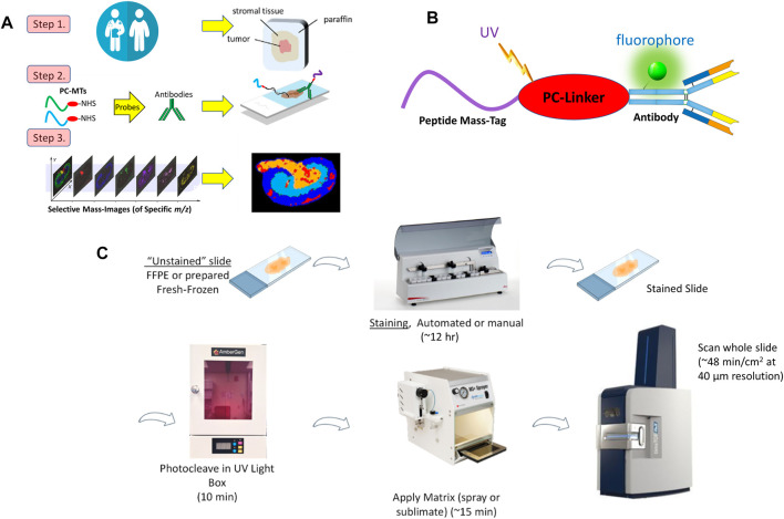
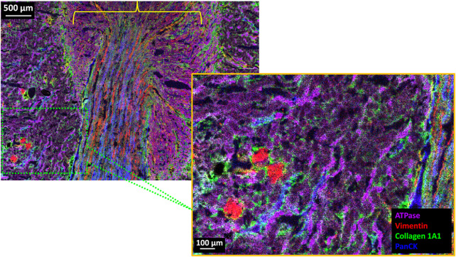
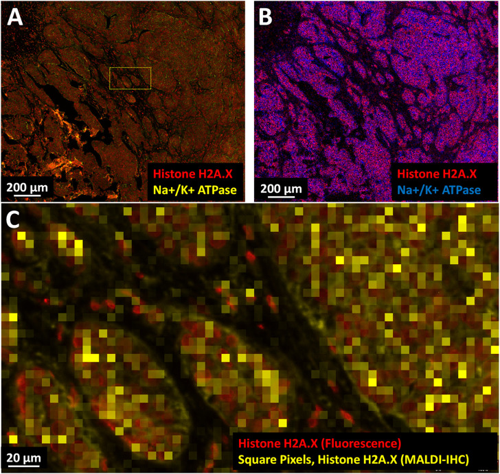
Matrix-assisted laser desorption/ionization mass spectrometry imaging (MALDI-MSI) is one of the most widely used methods for imaging the spatial distribution of unlabeled small molecules such as metabolites, lipids and drugs in tissues. Recent progress has enabled many improvements including the ability to achieve single cell spatial resolution, 3D-tissue image reconstruction, and the precise identification of different isomeric and isobaric molecules. However, MALDI-MSI of high molecular weight intact proteins in biospecimens has thus far been difficult to achieve. Conventional methods normally require proteolysis and peptide mass fingerprinting, have low spatial resolution, and typically detect only the most highly abundant proteins in an untargeted manner. In addition, MSI-based multiomic and multimodal workflows are needed which can image both small molecules and intact proteins from the same tissue. Such a capability can provide a more comprehensive understanding of the vast complexity of biological systems at the organ, tissue, and cellular levels of both normal and pathological function. A recently introduced top-down spatial imaging approach known as MALDI HiPLEX-IHC (MALDI-IHC for short) provides a basis for achieving this high-information content imaging of tissues and even individual cells. Based on novel photocleavable mass-tags conjugated to antibody probes, high-plex, multimodal and multiomic MALDI-based workflows have been developed to image both small molecules and intact proteins on the same tissue sample. Dual-labeled antibody probes enable multimodal mass spectrometry and fluorescent imaging of targeted intact proteins. A similar approach using the same photocleavable mass-tags can be applied to lectin and other probes. We detail here several examples of MALDI-IHC workflows designed to enable high-plex, multiomic and multimodal imaging of tissues at a spatial resolution as low as 5 µm. This approach is compared to other existing high-plex methods such as imaging mass cytometry, MIBI-TOF, GeoMx and CODEX. Finally, future applications of MALDI-IHC are discussed.
基质辅助激光解吸/电离质谱成像(MALDI-MSI)是用于成像组织中未标记小分子(如代谢物、脂质和药物)空间分布的最广泛使用的方法之一。最近的进展带来了许多改进,包括实现单细胞空间分辨率、三维组织图像重建以及精确鉴定不同的同分异构体和等压分子的能力。然而,生物样本中高分子量完整蛋白质的MALDI-MSI迄今为止一直难以实现。传统方法通常需要蛋白水解和肽质量指纹分析,空间分辨率低,并且通常以非靶向方式仅检测丰度最高的蛋白质。此外,还需要基于MSI的多组学和多模态工作流程,其能够对同一组织中的小分子和完整蛋白质进行成像。这种能力可以在正常和病理功能的器官、组织和细胞水平上更全面地了解生物系统的巨大复杂性。最近引入的一种自上而下的空间成像方法,称为MALDI HiPLEX-IHC(简称为MALDI-IHC),为实现这种组织甚至单个细胞的高信息含量成像提供了基础。基于与抗体探针偶联的新型光可裂解质量标签,已开发出高多重、多模态和多组学的基于MALDI的工作流程,以对同一组织样本上的小分子和完整蛋白质进行成像。双标记抗体探针能够对靶向完整蛋白质进行多模态质谱分析和荧光成像。使用相同光可裂解质量标签的类似方法可以应用于凝集素和其他探针。我们在此详细介绍几个MALDI-IHC工作流程的示例,这些工作流程旨在以低至5微米的空间分辨率对组织进行高多重、多组学和多模态成像。将这种方法与其他现有的高多重方法(如图像质谱流式细胞术、MIBI-TOF、GeoMx和CODEX)进行了比较。最后,讨论了MALDI-IHC的未来应用。