Chinnathambi Subashchandrabose, Das Rashmi
Neurobiology Group, Division of Biochemical Sciences, CSIR-National Chemical Laboratory, Dr. Homi Bhabha Road, Pune, 411008, India.
Academy of Scientific and Innovative Research (AcSIR), Ghaziabad, 201002, India.
Cell Biosci. 2023 May 23;13(1):95. doi: 10.1186/s13578-023-01028-0.
Tau protein forms neurofibrillary tangles and becomes deposited in the brain during Alzheimer's disease (AD). Tau oligomers are the most reactive species, mediating neurotoxic and inflammatory activity. Microglia are the immune cells in the central nervous system, sense the extracellular Tau via various cell surface receptors. Purinergic P2Y12 receptor can directly interact with Tau oligomers and mediates microglial chemotaxis via actin remodeling. The disease-associated microglia are associated with impaired migration and express a reduced level of P2Y12, but elevate the level of reactive oxygen species and pro-inflammatory cytokines.
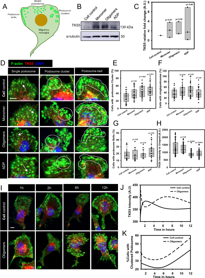
Here, we studied the formation and organization of various actin microstructures such as-podosome, filopodia and uropod in colocalization with actin nucleator protein Arp2 and scaffold protein TKS5 in Tau-induced microglia by fluorescence microscopy. Further, the relevance of P2Y12 signaling either by activation or blockage was studied in terms of actin structure formations and Tau deposits degradation by N9 microglia. Extracellular Tau oligomers facilitate the microglial migration via Arp2-associated podosome and filopodia formation through the involvement of P2Y12 signaling. Similarly, Tau oligomers induce the TKS5-associated podosome clustering in microglial lamella in a time-dependent manner. Moreover, the P2Y12 was evidenced to localize with F-actin-rich podosome and filopodia during Tau-deposit degradation. The blockage of P2Y12 signaling resulted in decreased microglial migration and Tau-deposit degradation.
The P2Y12 signaling mediate the formation of migratory actin structures like- podosome and filopodia to exhibit chemotaxis and degrade Tau deposit. These beneficial roles of P2Y12 in microglial chemotaxis, actin network remodeling and Tau clearance can be intervened as a therapeutic target in AD.
在阿尔茨海默病(AD)期间,tau蛋白形成神经原纤维缠结并沉积于大脑中。tau寡聚体是最具反应活性的物质,介导神经毒性和炎症活动。小胶质细胞是中枢神经系统中的免疫细胞,通过各种细胞表面受体感知细胞外tau蛋白。嘌呤能P2Y12受体可直接与tau寡聚体相互作用,并通过肌动蛋白重塑介导小胶质细胞趋化作用。疾病相关的小胶质细胞与迁移受损有关,P2Y12表达水平降低,但活性氧和促炎细胞因子水平升高。
在此,我们通过荧光显微镜研究了tau诱导的小胶质细胞中各种肌动蛋白微结构(如足体、丝状伪足和尾足)与肌动蛋白成核蛋白Arp2和支架蛋白TKS5共定位的形成和组织情况。此外,研究了通过激活或阻断P2Y12信号传导在N9小胶质细胞的肌动蛋白结构形成和tau沉积物降解方面的相关性。细胞外tau寡聚体通过P2Y12信号传导的参与,促进肌动蛋白相关的足体和丝状伪足形成,从而促进小胶质细胞迁移。同样,tau寡聚体以时间依赖性方式诱导小胶质细胞薄片中TKS5相关的足体聚集。此外,在tau沉积物降解过程中,P2Y12被证明定位于富含F-肌动蛋白的足体和丝状伪足。阻断P2Y12信号传导导致小胶质细胞迁移和tau沉积物降解减少。
P2Y12信号传导介导足体和丝状伪足等迁移性肌动蛋白结构的形成,以表现出趋化作用并降解tau沉积物。P2Y12在小胶质细胞趋化、肌动蛋白网络重塑和tau清除中的这些有益作用可作为AD的治疗靶点进行干预。