Institute for Pathology and Neuropathology, University Hospital Tübingen, Tübingen, Germany.
Transfusion Medicine, Medical Faculty of Tübingen, University of Tübingen, Tübingen, Germany; Centre for Clinical Transfusion Medicine Tübingen ZKT gGmbH, University of Tübingen, Tübingen, Germany.
Lab Invest. 2023 Aug;103(8):100179. doi: 10.1016/j.labinv.2023.100179. Epub 2023 May 22.
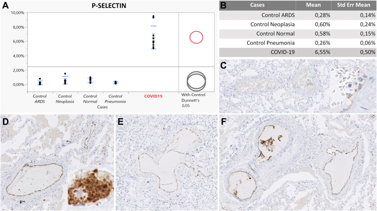
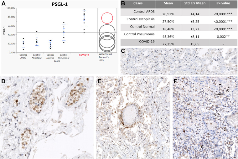
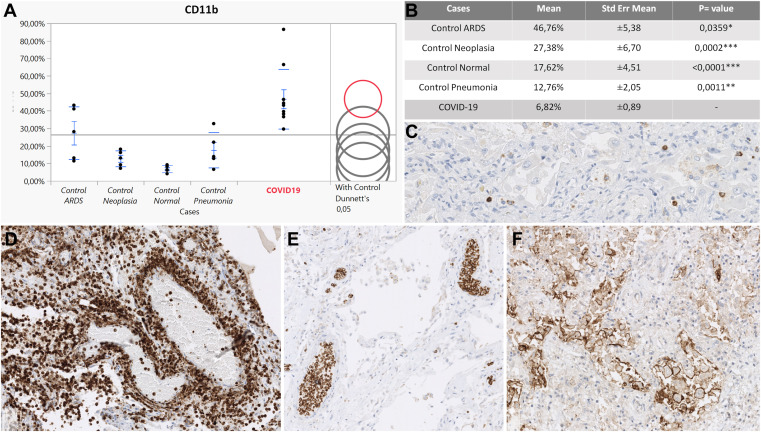
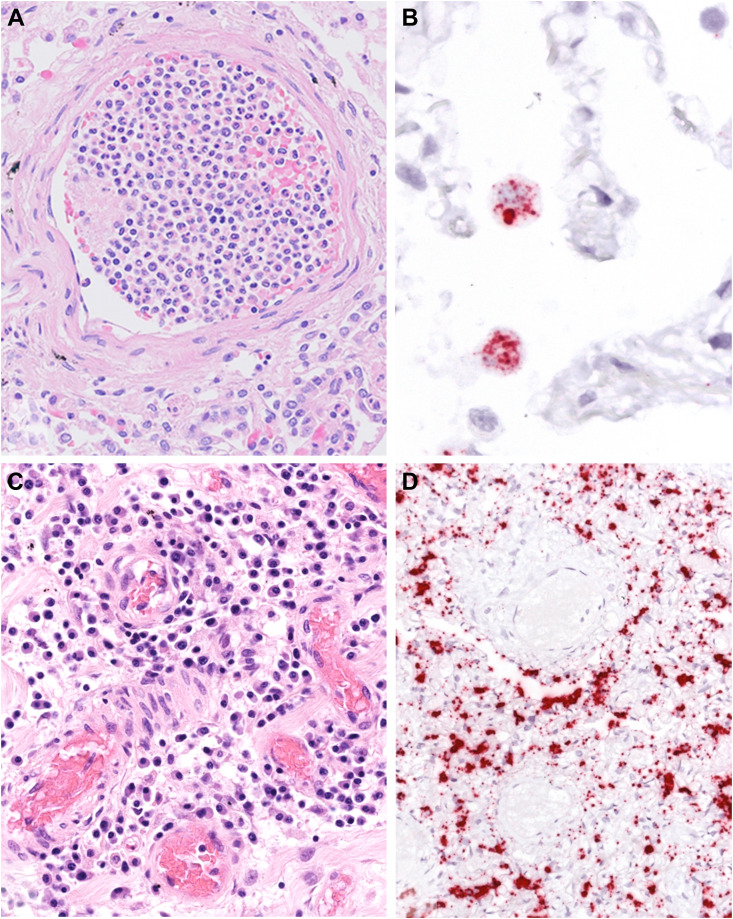
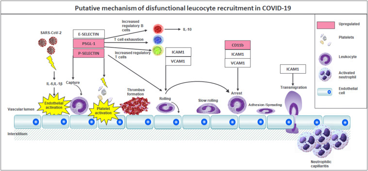
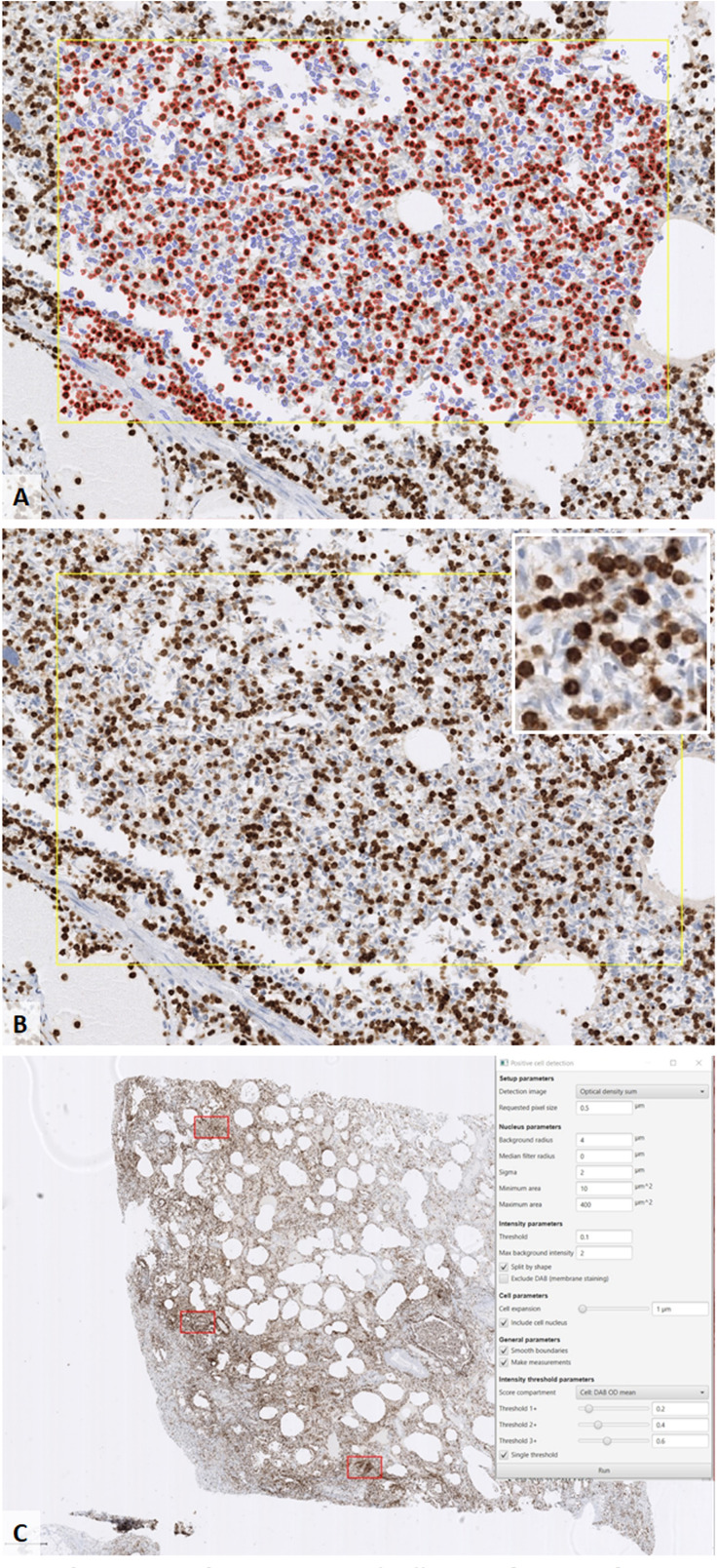
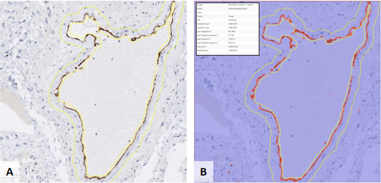
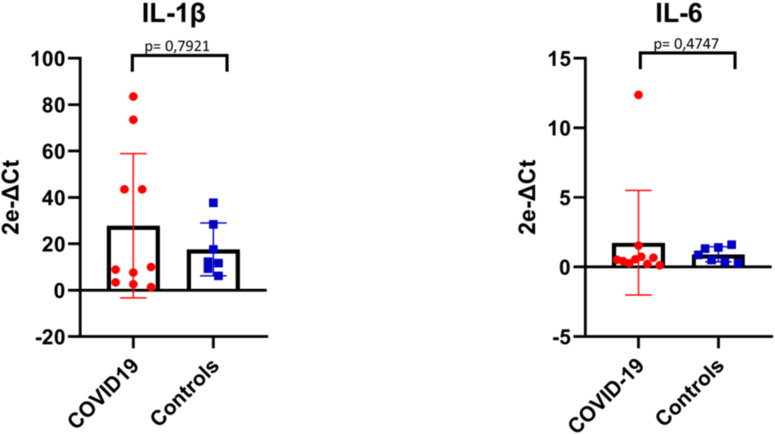
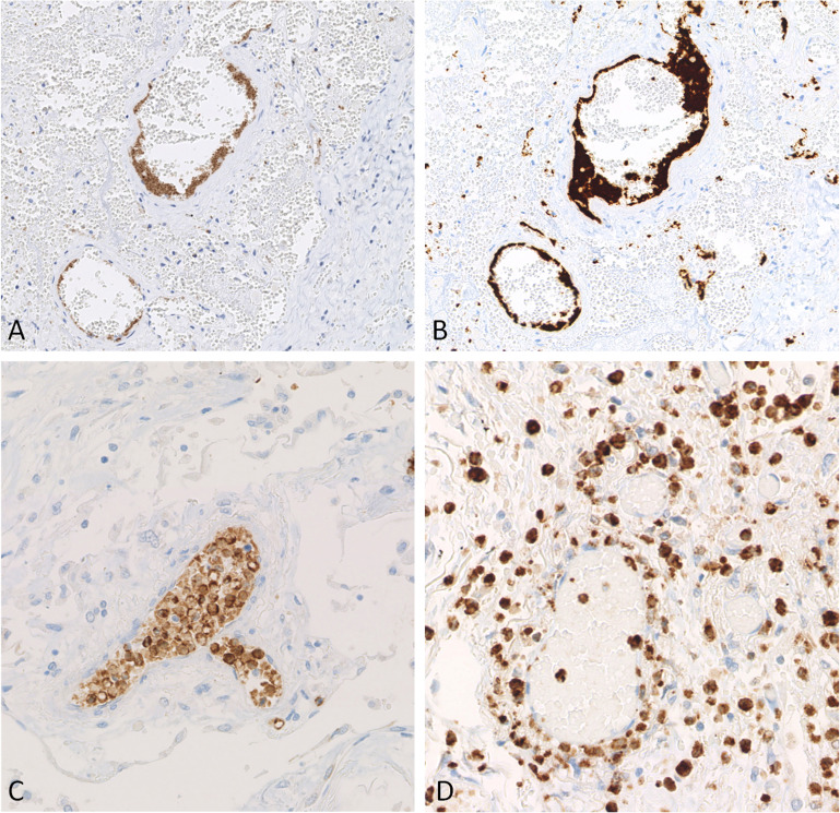
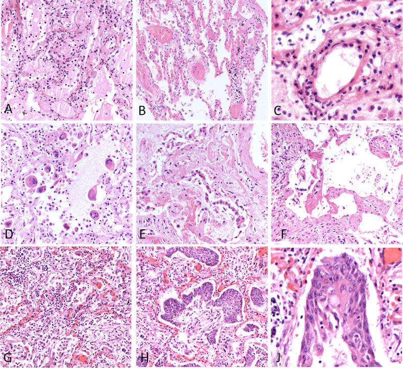
In critically ill patients infected with SARS-CoV-2, early leukocyte recruitment to the respiratory system was found to be orchestrated by leukocyte trafficking molecules accompanied by massive secretion of proinflammatory cytokines and hypercoagulability. Our study aimed to explore the interplay between leukocyte activation and pulmonary endothelium in different disease stages of fatal COVID-19. Our study comprised 10 COVID-19 postmortem lung specimens and 20 control lung samples (5 acute respiratory distress syndrome, 2 viral pneumonia, 3 bacterial pneumonia, and 10 normal), which were stained for antigens representing the different steps of leukocyte migration: E-selectin, P-selectin, PSGL-1, ICAM1, VCAM1, and CD11b. Image analysis software QuPath was used for quantification of positive leukocytes (PSGL-1 and CD11b) and endothelium (E-selectin, P-selectin, ICAM1, VCAM1). Expression of IL-6 and IL-1β was quantified by RT-qPCR. Expression of P-selectin and PSGL-1 was strongly increased in the COVID-19 cohort compared with all control groups (COVID-19:Controls, 17:23, P < .0001; COVID-19:Controls, 2:75, P < .0001, respectively). Importantly, P-selectin was found in endothelial cells and associated with aggregates of activated platelets adherent to the endothelial surface in COVID-19 cases. In addition, PSGL-1 staining disclosed positive perivascular leukocyte cuffs, reflecting capillaritis. Moreover, CD11b showed a strongly increased positivity in COVID-19 compared with all controls (COVID-19:Controls, 2:89; P = .0002), indicating a proinflammatory immune microenvironment. Of note, CD11b exhibited distinct staining patterns at different stages of COVID-19 disease. Only in cases with very short disease course, high levels of IL-1β and IL-6 mRNA were observed in lung tissue. The striking upregulation of PSGL-1 and P-selectin reflects the activation of this receptor-ligand pair in COVID-19, increasing the efficiency of initial leukocyte recruitment, thus promoting tissue damage and immunothrombosis. Our results show that endothelial activation and unbalanced leukocyte migration play a central role in COVID-19 involving the P-selectin-PSGL-1 axis.
在感染 SARS-CoV-2 的危重病患者中,早期白细胞向呼吸系统的募集被发现是由白细胞迁移分子协调的,伴随着大量促炎细胞因子的分泌和高凝状态。我们的研究旨在探讨白细胞激活与不同 COVID-19 致死阶段的肺内皮之间的相互作用。我们的研究包括 10 例 COVID-19 死后肺标本和 20 例对照肺标本(5 例急性呼吸窘迫综合征,2 例病毒性肺炎,3 例细菌性肺炎,10 例正常),这些标本用代表白细胞迁移不同步骤的抗原进行染色:E-选择素、P-选择素、PSGL-1、ICAM1、VCAM1 和 CD11b。使用图像分析软件 QuPath 对阳性白细胞(PSGL-1 和 CD11b)和内皮细胞(E-选择素、P-选择素、ICAM1、VCAM1)进行定量。通过 RT-qPCR 定量测定 IL-6 和 IL-1β 的表达。与所有对照组相比,COVID-19 组中 P-选择素和 PSGL-1 的表达均显著增加(COVID-19:对照组,17:23,P <.0001;COVID-19:对照组,2:75,P <.0001)。重要的是,在 COVID-19 病例中发现 P-选择素存在于内皮细胞中,并与附着在内皮表面的活化血小板聚集物相关。此外,PSGL-1 染色显示 COVID-19 中存在阳性血管周围白细胞袖套,反映出毛细血管炎。此外,与所有对照组相比,CD11b 在 COVID-19 中表达呈显著增加(COVID-19:对照组,2:89;P =.0002),表明存在促炎免疫微环境。值得注意的是,CD11b 在 COVID-19 疾病的不同阶段表现出不同的染色模式。仅在疾病病程极短的情况下,肺组织中观察到高水平的 IL-1β 和 IL-6 mRNA。PSGL-1 和 P-选择素的显著上调反映了 COVID-19 中该受体-配体对的激活,提高了初始白细胞募集的效率,从而促进组织损伤和免疫血栓形成。我们的结果表明,内皮细胞激活和白细胞迁移失衡在涉及 P-选择素-PSGL-1 轴的 COVID-19 中发挥核心作用。