Departments of Medicine (S.C., J.C., W.D., D.M.M., C.Y.), University of Rochester School of Medicine and Dentistry, Rochester, NY.
Now with Department of Cardiology, Ruijin Hospital (J.C.), Shanghai Jiao-Tong University School of Medicine, China.
Circ Res. 2023 Jul 7;133(2):138-157. doi: 10.1161/CIRCRESAHA.122.322264. Epub 2023 May 26.
Cyclic nucleotides play critical roles in cardiovascular biology and disease. PDE10A (phosphodiesterase 10A) is able to hydrolyze both cAMP and cGMP. PDE10A expression is induced in various human tumor cell lines, and PDE10A inhibition suppresses tumor cell growth. Chemotherapy drug such as doxorubicin (DOX) is widely used in chemotherapy. However, cardiotoxicity of DOX remains to be a serious clinical complication. In the current study, we aim to determine the role of PDE10A and the effect of PDE10A inhibition on cancer growth and cardiotoxicity induced by DOX.
We used global PDE10A knockout (KO) mice and PDE10A inhibitor TP-10 to block PDE10A function. DOX-induced cardiotoxicity was evaluated in C57Bl/6J mice and nude mice with implanted ovarian cancer xenografts. Isolated adult mouse cardiomyocytes and a human ovarian cancer cell line were used for in vitro functional and mechanistic studies.
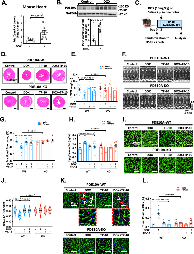
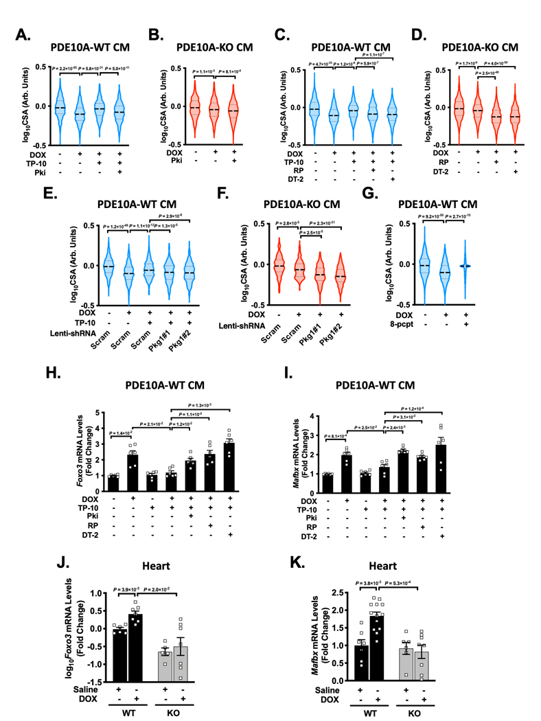
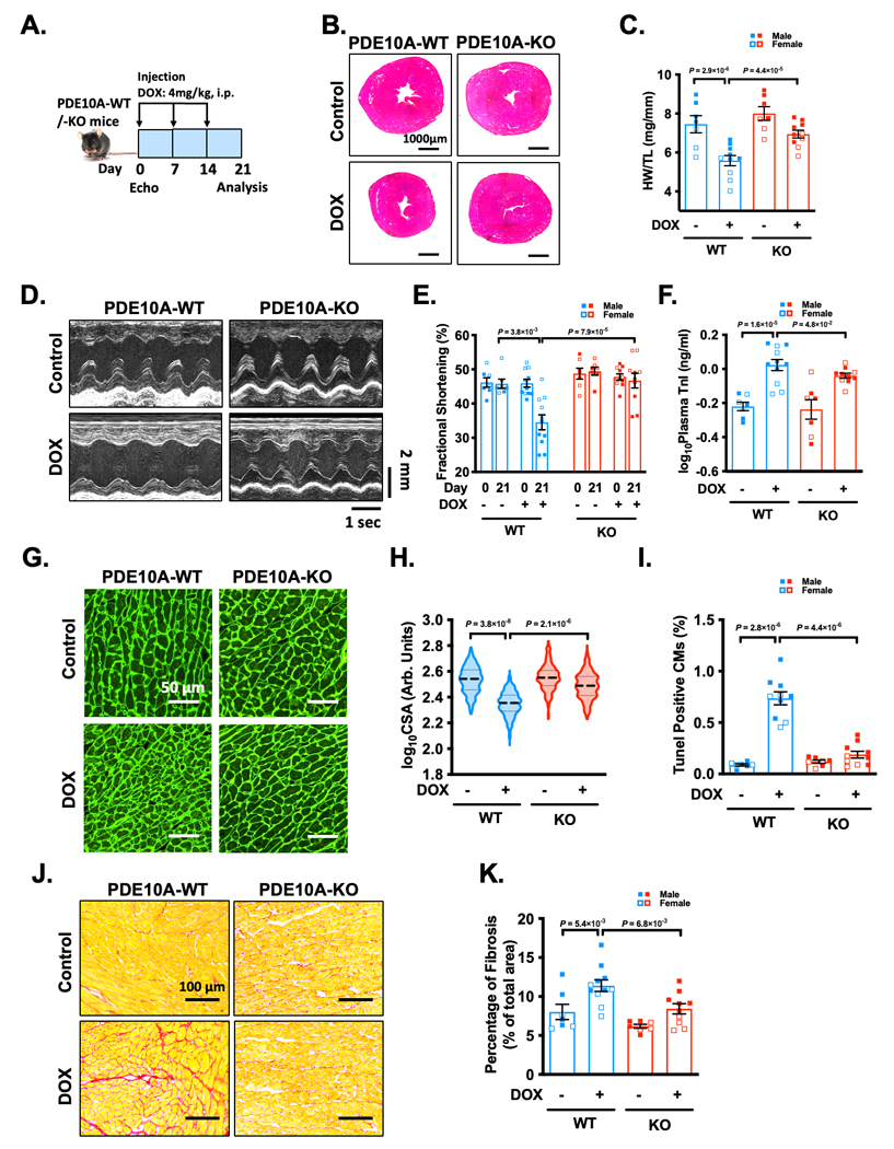
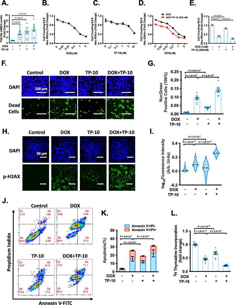
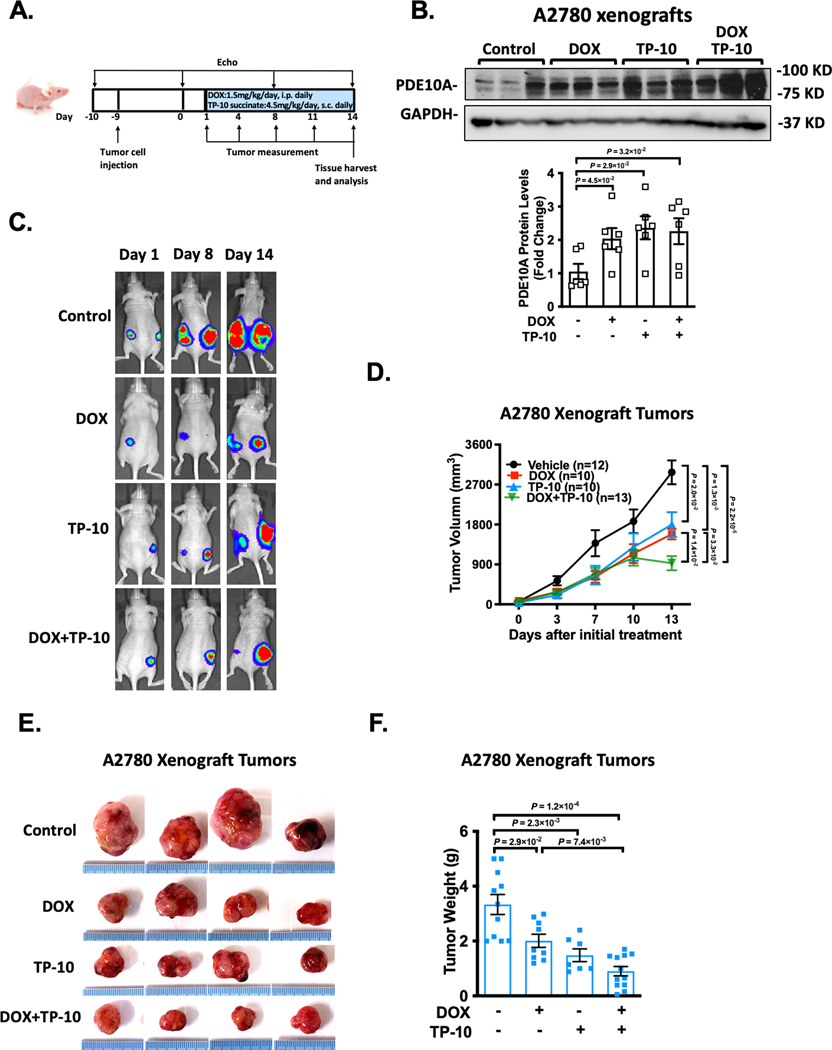
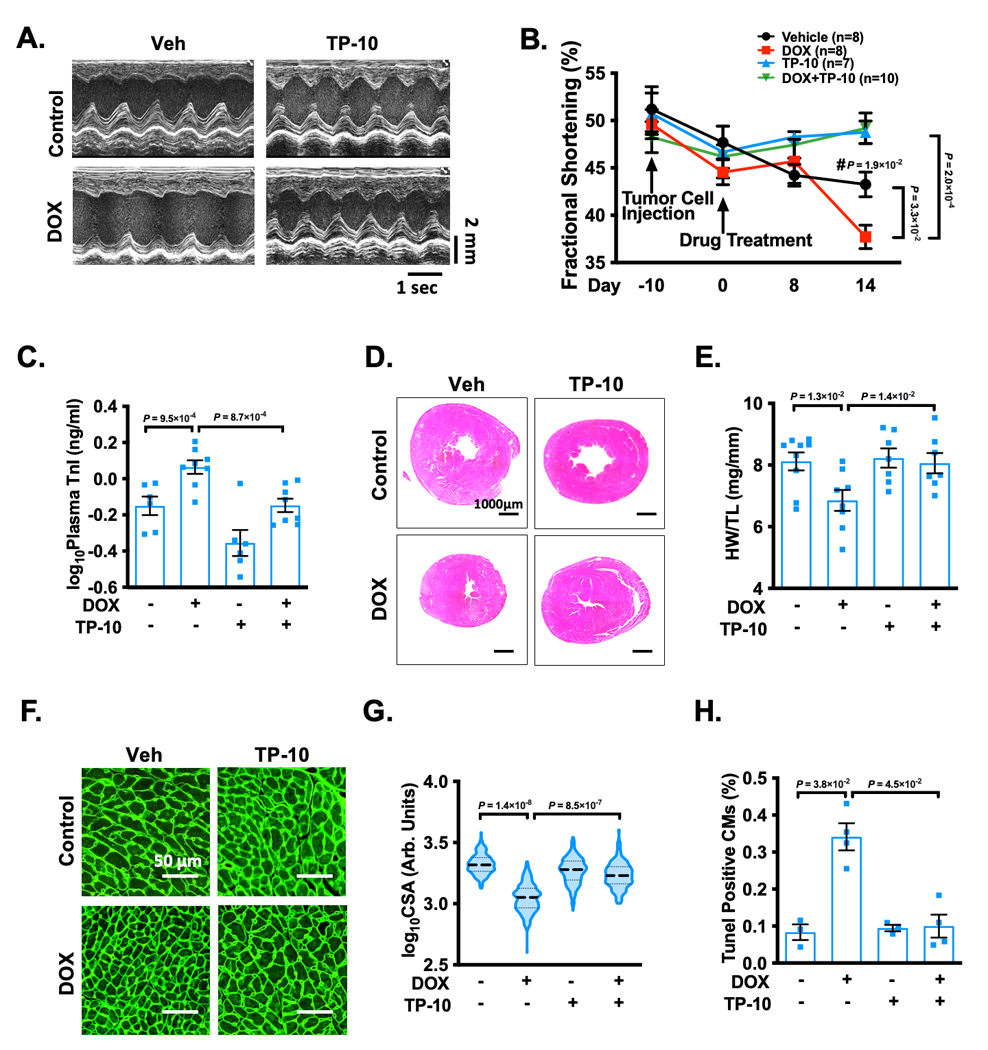
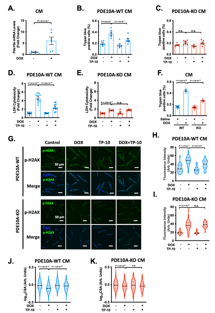
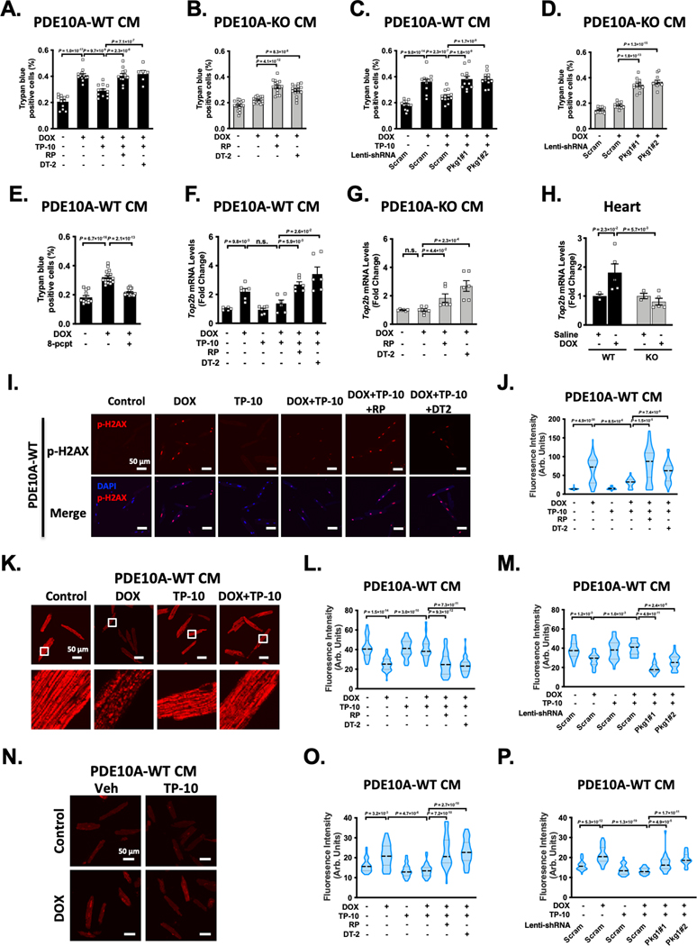
We found that PDE10A deficiency or inhibition alleviated DOX-induced myocardial atrophy, apoptosis, and dysfunction in C57Bl/6J mice. RNA sequencing study revealed a number of PDE10A-regulated signaling pathways involved in DOX-induced cardiotoxicity. PDE10A inhibition increased the death, decreased the proliferation, and potentiated the effect of DOX on various human cancer cells. Importantly, in nude mice with implanted ovarian cancer xenografts, PDE10A inhibition attenuated tumor growth while protecting DOX-induced cardiotoxicity. In isolated cardiomyocytes, PDE10A contributed to DOX-induced cardiomyocyte death via increasing Top2β (topoisomerase 2β) expression, mitochondrial dysfunction, and DNA damage by antagonizing cGMP/PKG (protein kinase G) signaling. PDE10A contributed to cardiomyocyte atrophy via potentiating FoxO3 (forkhead box O3) signaling via both cAMP/PKA (protein kinase A)- and cGMP/PKG-dependent signaling.
Taken together, our study elucidates a novel role for PDE10A in cardiotoxicity induced by DOX and cancer growth. Given that PDE10A has been already proven to be a safe drug target, PDE10A inhibition may represent a novel therapeutic strategy in cancer therapy, with effects preventing DOX-induced cardiotoxicity and simultaneously antagonizing cancer growth.
环核苷酸在心血管生物学和疾病中发挥着关键作用。PDE10A(磷酸二酯酶 10A)能够水解 cAMP 和 cGMP。各种人类肿瘤细胞系中均诱导表达 PDE10A,而 PDE10A 抑制可抑制肿瘤细胞生长。阿霉素(DOX)等化疗药物广泛用于化疗。然而,DOX 的心脏毒性仍然是一种严重的临床并发症。在本研究中,我们旨在确定 PDE10A 的作用以及 PDE10A 抑制对 DOX 诱导的肿瘤生长和心脏毒性的影响。
我们使用全局 PDE10A 敲除(KO)小鼠和 PDE10A 抑制剂 TP-10 来阻断 PDE10A 功能。在 C57Bl/6J 小鼠和植入卵巢癌异种移植的裸鼠中评估 DOX 诱导的心脏毒性。分离成年小鼠心肌细胞和人卵巢癌细胞系进行体外功能和机制研究。
我们发现,PDE10A 缺乏或抑制减轻了 C57Bl/6J 小鼠中 DOX 诱导的心肌萎缩、凋亡和功能障碍。RNA 测序研究揭示了许多 PDE10A 调节的信号通路参与 DOX 诱导的心脏毒性。PDE10A 抑制增加了各种人癌细胞的死亡、减少了增殖,并增强了 DOX 的作用。重要的是,在植入卵巢癌异种移植的裸鼠中,PDE10A 抑制减轻了肿瘤生长,同时保护了 DOX 诱导的心脏毒性。在分离的心肌细胞中,PDE10A 通过增加 Top2β(拓扑异构酶 2β)表达、线粒体功能障碍和 DNA 损伤来拮抗 cGMP/PKG(蛋白激酶 G)信号,导致 DOX 诱导的心肌细胞死亡。PDE10A 通过增强 FoxO3(叉头框 O3)信号来促进心肌细胞萎缩,该信号通过 cAMP/PKA(蛋白激酶 A)和 cGMP/PKG 依赖性信号传导。
总之,我们的研究阐明了 PDE10A 在 DOX 诱导的心脏毒性和肿瘤生长中的新作用。鉴于 PDE10A 已被证明是一种安全的药物靶点,PDE10A 抑制可能代表一种新的癌症治疗策略,具有预防 DOX 诱导的心脏毒性和同时拮抗肿瘤生长的作用。