• 文献检索
  • 文档翻译
  • 深度研究
  • 学术资讯
  • Suppr Zotero 插件Zotero 插件
  • 邀请有礼
  • 套餐&价格
  • 历史记录
应用&插件
Suppr Zotero 插件Zotero 插件浏览器插件Mac 客户端Windows 客户端微信小程序
定价
高级版会员购买积分包购买API积分包
服务
文献检索文档翻译深度研究API 文档MCP 服务
关于我们
关于 Suppr公司介绍联系我们用户协议隐私条款
关注我们

Suppr 超能文献

核心技术专利:CN118964589B侵权必究
粤ICP备2023148730 号-1Suppr @ 2026

文献检索

告别复杂PubMed语法,用中文像聊天一样搜索,搜遍4000万医学文献。AI智能推荐,让科研检索更轻松。

立即免费搜索

文件翻译

保留排版,准确专业,支持PDF/Word/PPT等文件格式,支持 12+语言互译。

免费翻译文档

深度研究

AI帮你快速写综述,25分钟生成高质量综述,智能提取关键信息,辅助科研写作。

立即免费体验

基于体积的杏仁核和海马亚区与记忆的关联:早期阿尔茨海默病的 3 年随访研究。

7T amygdala and hippocampus subfields in volumetry-based associations with memory: A 3-year follow-up study of early Alzheimer's disease.

机构信息

Charité - Universitätsmedizin Berlin, Corporate Member of Freie Universität Berlin and Humboldt-Universität zu Berlin, Department of Neurology, Charitéplatz 1, 10117 Berlin, Germany; Charité - Universitätsmedizin Berlin, Corporate Member of Freie Universität Berlin and Humboldt-Universität zu Berlin, NCRC - Neuroscience Clinical Research Center, Charitéplatz 1, 10117 Berlin, Germany.

Charité - Universitätsmedizin Berlin, Corporate Member of Freie Universität Berlin and Humboldt-Universität zu Berlin, Department of Neurology, Charitéplatz 1, 10117 Berlin, Germany; Charité - Universitätsmedizin Berlin, Corporate Member of Freie Universität Berlin and Humboldt-Universität zu Berlin, NCRC - Neuroscience Clinical Research Center, Charitéplatz 1, 10117 Berlin, Germany.

出版信息

Neuroimage Clin. 2023;38:103439. doi: 10.1016/j.nicl.2023.103439. Epub 2023 May 24.

DOI:10.1016/j.nicl.2023.103439
PMID:37253284
原文链接:https://pmc.ncbi.nlm.nih.gov/articles/PMC10236463/
Abstract

INTRODUCTION

The hippocampus is the most prominent single region of interest (ROI) for the diagnosis and prediction of Alzheimer's disease (AD). However, its suitability in the earliest stages of cognitive decline, i.e., subjective cognitive decline (SCD), remains uncertain which warrants the pursuit of alternative or complementary regions. The amygdala might be a promising candidate, given its implication in memory as well as other psychiatric disorders, e.g. depression and anxiety, which are prevalent in SCD. In this 7 tesla (T) magnetic resonance imaging (MRI) study, we aimed to compare the contribution of volumetric measurements of the hippocampus, the amygdala, and their respective subfields, for early diagnosis and prediction in an AD-related study population.

METHODS

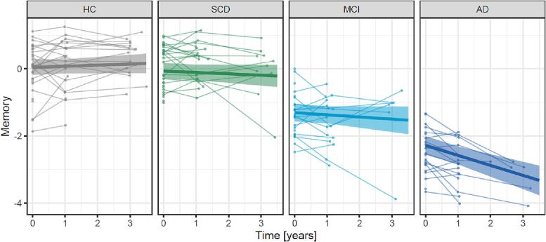
Participants from a longitudinal study were grouped into SCD (n = 29), mild cognitive impairment (MCI, n = 23), AD (n = 22) and healthy control (HC, n = 31). All participants underwent 7T MRI at baseline and extensive neuropsychological testing at up to three visits (baseline n = 105, 1-year n = 78, 3-year n = 39). Analysis of covariance (ANCOVA) was used to assess group differences of baseline volumes of the amygdala and the hippocampus and their subfields. Linear mixed models were used to estimate the effects of baseline volumes on yearly changes of a z-scaled memory score. All models were adjusted to age, sex and education.

RESULTS

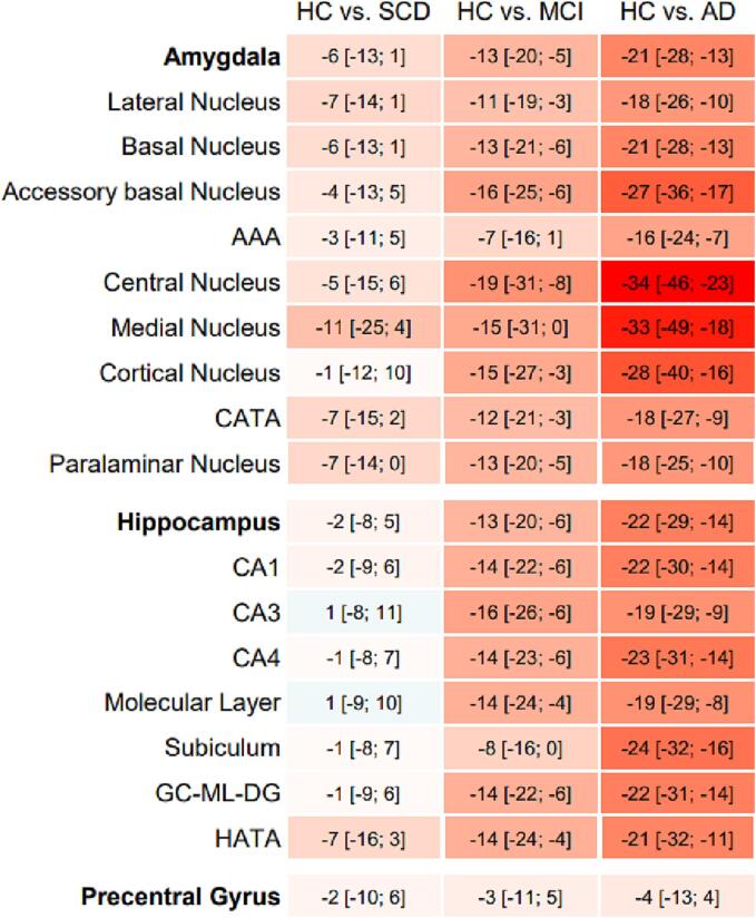
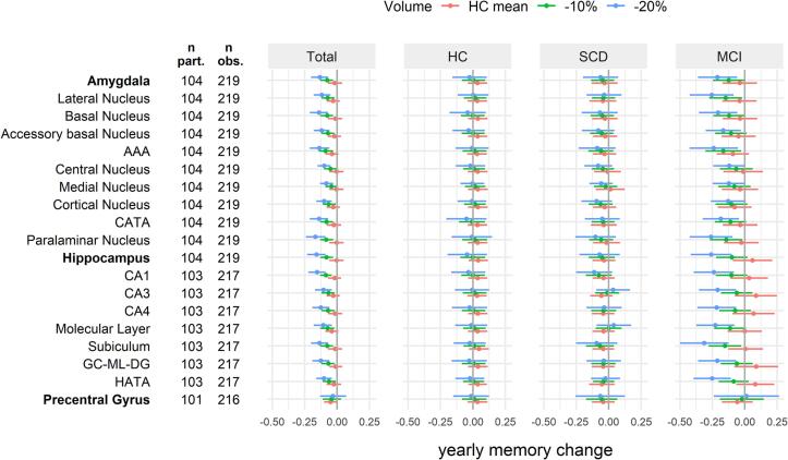
Compared to the HC group, individuals with SCD showed smaller amygdala ROI volumes (range across subfields -11% to -1%), but not hippocampus ROI volumes (-2% to 1%) except for the hippocampus-amygdala-transition-area (-7%). However, cross-sectional associations between baseline memory and volumes were smaller for amygdala ROIs (std. ß [95% CI] ranging between 0.16 [0.08; 0.25] and 0.46 [0.31; 0.60]) than hippocampus ROIs (between 0.32 [0.19; 0.44] and 0.53 [0.40; 0.67]). Further, the association of baseline volumes with yearly memory change in the HC and SCD groups was similarly weak for amygdala ROIs and hippocampus ROIs. In the MCI group, volumes of amygdala ROIs were associated with a relevant yearly memory decline [95% CI] ranging between -0.12 [-0.24; 0.00] and -0.26 [-0.42; -0.09] for individuals with 20% smaller volumes than the HC group. However, effects were stronger for hippocampus ROIs with a corresponding yearly memory decline ranging between -0.21 [-0.35; -0.07] and -0.31 [-0.50; -0.13].

CONCLUSION

Volumes of amygdala ROIs, as determined by 7T MRI, might contribute to objectively and non-invasively identify patients with SCD, and thus aid early diagnosis and treatment of individuals at risk to develop dementia due to AD, however associations with other psychiatric disorders should be evaluated in further studies. The amygdala's value in the prediction of longitudinal memory changes in the SCD group remains questionable. Primarily in patients with MCI, memory decline over 3 years appears to be more strongly associated with volumes of hippocampus ROIs than amygdala ROIs.

摘要

简介

海马体是诊断和预测阿尔茨海默病(AD)最突出的单一感兴趣区(ROI)。然而,它在认知能力下降的早期阶段,即主观认知下降(SCD)的适用性尚不确定,这需要寻找替代或补充的区域。杏仁核可能是一个很有前途的候选者,因为它在记忆以及其他精神疾病,如抑郁症和焦虑症,都有一定的关联,而这些疾病在 SCD 中很常见。在这项 7 特斯拉(T)磁共振成像(MRI)研究中,我们旨在比较海马体、杏仁核及其各自的亚区的容积测量在 AD 相关研究人群中的早期诊断和预测中的贡献。

方法

来自一项纵向研究的参与者被分为 SCD(n=29)、轻度认知障碍(MCI,n=23)、AD(n=22)和健康对照组(HC,n=31)。所有参与者在基线时进行 7T MRI 检查,并在最多三次访视时进行广泛的神经心理学测试(基线 n=105,1 年 n=78,3 年 n=39)。协方差分析(ANCOVA)用于评估基线时杏仁核和海马体及其亚区的组间体积差异。线性混合模型用于估计基线体积对 z 标准化记忆评分的年度变化的影响。所有模型均调整为年龄、性别和教育程度。

结果

与 HC 组相比,SCD 患者的杏仁核 ROI 体积较小(各亚区范围为-11%至-1%),但海马体 ROI 体积无差异(-2%至 1%),除了海马体-杏仁核过渡区(-7%)。然而,与海马体 ROI 相比,基线记忆与体积之间的横断面相关性较小(标准β[95%CI]范围在 0.16[0.08;0.25]和 0.46[0.31;0.60]之间)。进一步的,在 HC 和 SCD 组中,基线体积与每年记忆变化的相关性对于杏仁核 ROI 和海马体 ROI 同样较弱。在 MCI 组中,杏仁核 ROI 的体积与相关的每年记忆下降相关[95%CI]范围在-0.12[-0.24;0.00]和-0.26[-0.42;0.09],与 HC 组相比,个体的体积减少了 20%。然而,对于海马体 ROI 来说,其影响更强,相应的每年记忆下降范围在-0.21[-0.35;0.07]和-0.31[-0.50;0.13]之间。

结论

7T MRI 确定的杏仁核 ROI 体积可能有助于客观、无创地识别 SCD 患者,从而有助于早期诊断和治疗 AD 风险人群,但应在进一步研究中评估与其他精神疾病的关联。杏仁核在 SCD 组的纵向记忆变化预测中的价值仍存在疑问。主要在 MCI 患者中,3 年内的记忆下降与海马体 ROI 体积的相关性明显强于杏仁核 ROI 体积。

https://cdn.ncbi.nlm.nih.gov/pmc/blobs/4eaf/10236463/7d2e69324454/gr3.jpg
https://cdn.ncbi.nlm.nih.gov/pmc/blobs/4eaf/10236463/a77dd618b5a1/gr1.jpg
https://cdn.ncbi.nlm.nih.gov/pmc/blobs/4eaf/10236463/4c1fb26cff24/gr2.jpg
https://cdn.ncbi.nlm.nih.gov/pmc/blobs/4eaf/10236463/7d2e69324454/gr3.jpg
https://cdn.ncbi.nlm.nih.gov/pmc/blobs/4eaf/10236463/a77dd618b5a1/gr1.jpg
https://cdn.ncbi.nlm.nih.gov/pmc/blobs/4eaf/10236463/4c1fb26cff24/gr2.jpg
https://cdn.ncbi.nlm.nih.gov/pmc/blobs/4eaf/10236463/7d2e69324454/gr3.jpg

相似文献

1
7T amygdala and hippocampus subfields in volumetry-based associations with memory: A 3-year follow-up study of early Alzheimer's disease.基于体积的杏仁核和海马亚区与记忆的关联:早期阿尔茨海默病的 3 年随访研究。
Neuroimage Clin. 2023;38:103439. doi: 10.1016/j.nicl.2023.103439. Epub 2023 May 24.
2
Volume changes of hippocampal and amygdala subfields in patients with mild cognitive impairment and Alzheimer's disease.海马和杏仁核亚区体积变化在轻度认知障碍和阿尔茨海默病患者中的研究。
Acta Neurol Belg. 2023 Aug;123(4):1381-1393. doi: 10.1007/s13760-023-02235-9. Epub 2023 Apr 12.
3
Smaller grey matter volume in the central olfactory system in mild cognitive impairment.轻度认知障碍患者的中央嗅觉系统灰质体积较小。
Exp Gerontol. 2023 Nov;183:112325. doi: 10.1016/j.exger.2023.112325. Epub 2023 Nov 11.
4
Structural Alteration of Medial Temporal Lobe Subfield in the Amnestic Mild Cognitive Impairment Stage of Alzheimer's Disease.内侧颞叶亚区结构改变在阿尔茨海默病遗忘型轻度认知障碍阶段。
Neural Plast. 2022 Jan 24;2022:8461235. doi: 10.1155/2022/8461235. eCollection 2022.
5
Structural magnetic resonance imaging for the early diagnosis of dementia due to Alzheimer's disease in people with mild cognitive impairment.用于轻度认知障碍患者阿尔茨海默病所致痴呆早期诊断的结构磁共振成像
Cochrane Database Syst Rev. 2020 Mar 2;3(3):CD009628. doi: 10.1002/14651858.CD009628.pub2.
6
Atrophy of specific amygdala subfields in subjects converting to mild cognitive impairment.向轻度认知障碍转化的受试者中特定杏仁核亚区的萎缩
Alzheimers Dement (N Y). 2023 Dec 3;9(4):e12436. doi: 10.1002/trc2.12436. eCollection 2023 Oct-Dec.
7
Cross-sectional and longitudinal characterization of SCD patients recruited from the community versus from a memory clinic: subjective cognitive decline, psychoaffective factors, cognitive performances, and atrophy progression over time.从社区和记忆诊所招募的 SCD 患者的横断面和纵向特征:主观认知下降、心理情感因素、认知表现以及随时间的进展的萎缩。
Alzheimers Res Ther. 2019 Jul 8;11(1):61. doi: 10.1186/s13195-019-0514-z.
8
Association between fully automated MRI-based volumetry of different brain regions and neuropsychological test performance in patients with amnestic mild cognitive impairment and Alzheimer's disease.基于全自动化 MRI 的不同脑区容积测量与遗忘型轻度认知障碍和阿尔茨海默病患者神经心理学测试表现的相关性。
Eur Arch Psychiatry Clin Neurosci. 2013 Jun;263(4):335-44. doi: 10.1007/s00406-012-0350-7. Epub 2012 Sep 1.
9
Memory impairment in Amyloidβ-status Alzheimer's disease is associated with a reduction in CA1 and dentate gyrus volume: In vivo MRI at 7T.阿尔茨海默病患者淀粉样蛋白β状态下的记忆障碍与 CA1 和齿状回体积减少有关:7T 体内 MRI。
Neuroimage. 2024 Apr 15;292:120607. doi: 10.1016/j.neuroimage.2024.120607. Epub 2024 Apr 12.
10
Asymmetry of Hippocampus and Amygdala Defect in Subjective Cognitive Decline Among the Community Dwelling Chinese.中国社区居民主观认知下降中海马体和杏仁核缺陷的不对称性
Front Psychiatry. 2018 Jun 11;9:226. doi: 10.3389/fpsyt.2018.00226. eCollection 2018.

引用本文的文献

1
Implicating neuroinflammation in hippocampus, prefrontal cortex and amygdala with cognitive deficit: a narrative review.海马体、前额叶皮质和杏仁核中的神经炎症与认知缺陷的关联:一项叙述性综述。
3 Biotech. 2025 Sep;15(9):320. doi: 10.1007/s13205-025-04468-2. Epub 2025 Aug 30.
2
Development of a Predictive Model for the Progression of Subjective Cognitive Decline: A Longitudinal Study.主观认知衰退进展预测模型的开发:一项纵向研究。
Brain Behav. 2025 Aug;15(8):e70719. doi: 10.1002/brb3.70719.
3
A perspective on the mechanisms of herbal medicine for cognitive impairment.
关于草药治疗认知障碍机制的观点。
Front Neurol. 2025 Jul 4;16:1610542. doi: 10.3389/fneur.2025.1610542. eCollection 2025.
4
Validation of dementia-free status in the iranian brain imaging database using ADNI data: a transition point from middle age to older adulthood.利用阿尔茨海默病神经影像学计划(ADNI)数据验证伊朗脑成像数据库中的无痴呆状态:从中年到老年的过渡点。
BMC Neurol. 2025 Jul 10;25(1):286. doi: 10.1186/s12883-025-04293-3.
5
A Comparison of the Brain Parameters of Thais with Normal Cognition, Mild Cognitive Impairment, and Dementia.认知正常、轻度认知障碍和痴呆的泰国人的脑参数比较。
Brain Sci. 2025 Jan 23;15(2):105. doi: 10.3390/brainsci15020105.
6
Plasma p-tau181 and GFAP reflect 7T MR-derived changes in Alzheimer's disease: A longitudinal study of structural and functional MRI and MRS.血浆p-tau181和胶质纤维酸性蛋白反映了7T磁共振成像在阿尔茨海默病中的变化:一项结构和功能磁共振成像及磁共振波谱的纵向研究
Alzheimers Dement. 2024 Dec;20(12):8684-8699. doi: 10.1002/alz.14318. Epub 2024 Nov 19.
7
Explainable early detection of Alzheimer's disease using ROIs and an ensemble of 138 3D vision transformers.使用 ROI 和 138 个 3D 视觉转换器的集合进行可解释的阿尔茨海默病早期检测。
Sci Rep. 2024 Nov 12;14(1):27756. doi: 10.1038/s41598-024-76313-0.
8
Structural disruption in subjective cognitive decline and mild cognitive impairment.主观认知下降和轻度认知障碍中的结构破坏。
Brain Imaging Behav. 2024 Dec;18(6):1536-1548. doi: 10.1007/s11682-024-00933-3. Epub 2024 Oct 7.
9
Alteration in amygdala subfield volumes and their association with cognition in mild cognitive impairment.轻度认知障碍患者杏仁核亚区体积改变及其与认知的关联
J Neurol. 2024 Aug;271(8):5460-5467. doi: 10.1007/s00415-024-12500-3. Epub 2024 Jun 15.
10
Atrophy of hippocampal subfields and amygdala nuclei in subjects with mild cognitive impairment progressing to Alzheimer's disease.轻度认知障碍进展为阿尔茨海默病患者海马亚区和杏仁核萎缩
Heliyon. 2024 Mar 8;10(6):e27429. doi: 10.1016/j.heliyon.2024.e27429. eCollection 2024 Mar 30.