Zhu Qin, Barnes Clayton E, Mannes Philip Z, Latoche Joseph D, Day Kathryn E, Nedrow Jessie R, Novelli Enrico M, Anderson Carolyn J, Tavakoli Sina
Department of Radiology, University of Pittsburgh, UPMC Presbyterian Hospital, 200 Lothrop Street, Suite E200, Pittsburgh, PA, 15213, USA.
Medical Scientist Training Program, University of Pittsburgh, Pittsburgh, PA, USA.
EJNMMI Res. 2023 Jun 5;13(1):55. doi: 10.1186/s13550-023-01006-0.
The lack of noninvasive methods for assessment of dysregulated inflammation as a major driver of fibrosis (i.e., inflammation-fibrosis axis) has been a major challenge to precision management of fibrotic lung diseases. Here, we determined the potential of very late antigen-4 (VLA-4)-targeted positron emission tomography (PET) to detect inflammation in a mouse model of bleomycin-induced fibrotic lung injury.
Single time-point and longitudinal VLA-4-targeted PET was performed using a high-affinity peptidomimetic radiotracer, Cu-LLP2A, at weeks 1, 2, and 4 after bleomycin-induced (2.5 units/kg) lung injury in C57BL/6J mice. The severity of fibrosis was determined by measuring the hydroxyproline content of the lungs and expression of markers of extracellular matrix remodeling. Flow cytometry and histology was performed to determine VLA-4 expression across different leukocyte subsets and their spatial distribution.
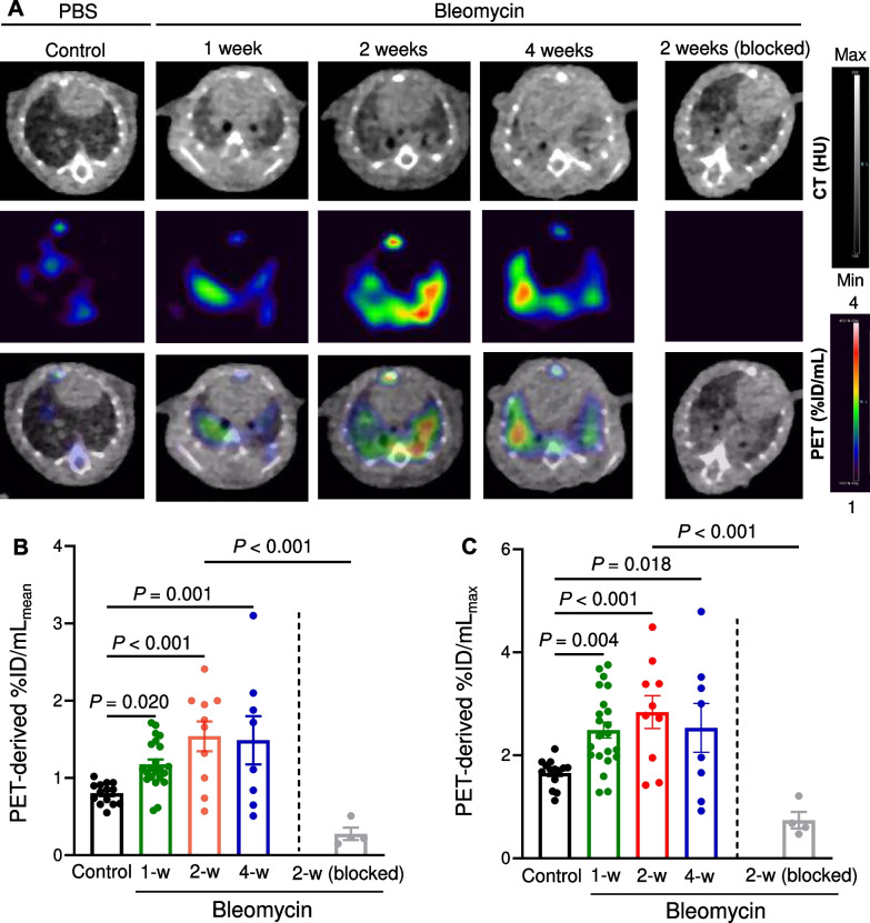
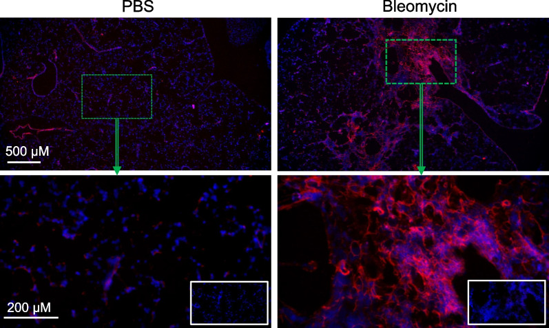
Lung uptake of Cu-LLP2A was significantly elevated throughout different stages of the progression of bleomycin-induced injury. High lung uptake of Cu-LLP2A at week-1 post-bleomycin was a predictor of poor survival over the 4-week follow up, supporting the prognostic potential of Cu-LLP2A PET during the early stage of the disease. Additionally, the progressive increase in Cu-LLP2A uptake from week-1 to week-4 post-bleomycin correlated with the ultimate extent of lung fibrosis and ECM remodeling. Flow cytometry revealed that LLP2A binding was restricted to leukocytes. A combination of increased expression of VLA-4 by alveolar macrophages and accumulation of VLA-4-expressing interstitial and monocyte-derived macrophages as well as dendritic cells was noted in bleomycin-injured, compared to control, lungs. Histology confirmed the increased expression of VLA-4 in bleomycin-injured lungs, particularly in inflamed and fibrotic regions.
VLA-4-targeted PET allows for assessment of the inflammation-fibrosis axis and prediction of disease progression in a murine model. The potential of Cu-LLP2A PET for assessment of the inflammation-fibrosis axis in human fibrotic lung diseases needs to be further investigated.
缺乏用于评估失调炎症(作为纤维化的主要驱动因素,即炎症 - 纤维化轴)的非侵入性方法,一直是纤维化肺病精准管理的重大挑战。在此,我们确定了靶向极迟抗原 - 4(VLA - 4)的正电子发射断层扫描(PET)在博来霉素诱导的纤维化肺损伤小鼠模型中检测炎症的潜力。
在C57BL / 6J小鼠中,于博来霉素诱导(2.5单位/千克)肺损伤后的第1、2和4周,使用高亲和力拟肽放射性示踪剂Cu - LLP2A进行单时间点和纵向VLA - 4靶向PET检查。通过测量肺组织中的羟脯氨酸含量和细胞外基质重塑标志物的表达来确定纤维化的严重程度。进行流式细胞术和组织学检查,以确定不同白细胞亚群中VLA - 4的表达及其空间分布。
在博来霉素诱导损伤的不同进展阶段,肺对Cu - LLP2A的摄取均显著升高。博来霉素注射后第1周肺对Cu - LLP2A的高摄取是4周随访期间生存率低的一个预测指标,这支持了Cu - LLP2A PET在疾病早期的预后潜力。此外,博来霉素注射后第1周到第4周Cu - LLP2A摄取的逐渐增加与肺纤维化和细胞外基质重塑的最终程度相关。流式细胞术显示LLP2A结合仅限于白细胞。与对照肺相比,在博来霉素损伤的肺中,观察到肺泡巨噬细胞VLA - 4表达增加,以及表达VLA - 4的间质和单核细胞衍生的巨噬细胞以及树突状细胞的积累。组织学证实博来霉素损伤的肺中VLA - 4表达增加,特别是在炎症和纤维化区域。
靶向VLA - 4的PET能够评估小鼠模型中的炎症 - 纤维化轴并预测疾病进展。Cu - LLP2A PET在人类纤维化肺病中评估炎症 - 纤维化轴的潜力需要进一步研究。