• 文献检索
  • 文档翻译
  • 深度研究
  • 学术资讯
  • Suppr Zotero 插件Zotero 插件
  • 邀请有礼
  • 套餐&价格
  • 历史记录
应用&插件
Suppr Zotero 插件Zotero 插件浏览器插件Mac 客户端Windows 客户端微信小程序
定价
高级版会员购买积分包购买API积分包
服务
文献检索文档翻译深度研究API 文档MCP 服务
关于我们
关于 Suppr公司介绍联系我们用户协议隐私条款
关注我们

Suppr 超能文献

核心技术专利:CN118964589B侵权必究
粤ICP备2023148730 号-1Suppr @ 2026

文献检索

告别复杂PubMed语法,用中文像聊天一样搜索,搜遍4000万医学文献。AI智能推荐,让科研检索更轻松。

立即免费搜索

文件翻译

保留排版,准确专业,支持PDF/Word/PPT等文件格式,支持 12+语言互译。

免费翻译文档

深度研究

AI帮你快速写综述,25分钟生成高质量综述,智能提取关键信息,辅助科研写作。

立即免费体验

扩张型心肌病患者的固有 T1 高区域和左心室射血分数恢复。

Native T1 high region and left ventricular ejection fraction recovery in patients with dilated cardiomyopathy.

机构信息

Department of Cardiovascular Medicine, Kitasato University School of Medicine, 1-15-1 Kitasato, Minami-ku, Sagamihara, Kanagawa, 252-0329, Japan.

Department of Cardiology, Leiden University Medical Center, Leiden, The Netherlands.

出版信息

Int J Cardiovasc Imaging. 2023 Sep;39(9):1785-1793. doi: 10.1007/s10554-023-02888-w. Epub 2023 Jun 8.

DOI:10.1007/s10554-023-02888-w
PMID:37291379
原文链接:https://pmc.ncbi.nlm.nih.gov/articles/PMC10520178/
Abstract

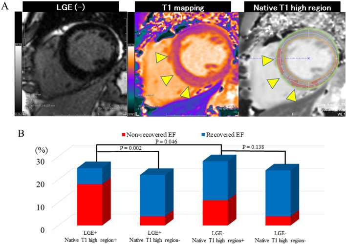
Native T1 mapping is used to assess myocardial tissue characteristics without gadolinium contrast agents. The focal T1 high-intensity region can indicate myocardial alterations. This study aimed to identify the association between the native T1 mapping including the native T1 high region and left ventricular ejection fraction (LVEF) recovery in patients with dilated cardiomyopathy (DCM). Patients with newly diagnosed DCM (LVEF of < 45%) who underwent cardiac magnetic resonance imaging with native T1 mapping were included in the analysis. Native T1 high region was defined as a signal intensity of > 5 SD in the remote myocardium. Recovered EF was defined as a follow-up LVEF of ≥ 45% and an LVEF increase of ≥ 10% after 2 years from baseline. Seventy-one patients met the inclusion criteria for this study. Forty-four patients (61.9%) achieved recovered EF. Logistic regression analysis showed that the native T1 value (OR: 0.98; 95% CI: 0.96-0.99; P = 0.014) and the native T1 high region (OR: 0.17; 95% CI: 0.05-0.55; P = 0.002), but not late gadolinium enhancement, were independent predictors of recovered EF. Compared with native T1 value alone, combined native T1 high region and native T1 value improved the area under the curve from 0.703 to 0.788 for predicting recovered EF. Myocardial damage, which was quantified using native T1 mapping and the native T1 high region were independently associated with recovered EF in patients with newly diagnosed DCM.

摘要

心肌组织的固有 T1 mapping 可用于评估心肌组织特征,而无需使用钆对比剂。局灶性 T1 高强度区域可提示心肌改变。本研究旨在确定扩张型心肌病(DCM)患者的固有 T1 映射(包括固有 T1 高区域)与左心室射血分数(LVEF)恢复之间的相关性。本研究纳入了经心脏磁共振成像固有 T1 映射检查的新发 DCM 患者(LVEF < 45%)。固有 T1 高区域定义为远场心肌信号强度> 5 个标准差。恢复的 EF 定义为随访时 LVEF ≥ 45%,且与基线相比 2 年后 LVEF 增加≥ 10%。71 名患者符合本研究纳入标准。44 名患者(61.9%)达到恢复 EF。Logistic 回归分析显示,固有 T1 值(OR:0.98;95%CI:0.96-0.99;P=0.014)和固有 T1 高区域(OR:0.17;95%CI:0.05-0.55;P=0.002),而不是晚期钆增强,是恢复 EF 的独立预测因子。与固有 T1 值相比,固有 T1 高区域和固有 T1 值联合应用可提高预测恢复 EF 的曲线下面积,从 0.703 提高到 0.788。心肌损伤是通过固有 T1 映射和固有 T1 高区域定量评估的,与新发 DCM 患者的恢复 EF 独立相关。

https://cdn.ncbi.nlm.nih.gov/pmc/blobs/dac5/10520178/91957e2311d0/10554_2023_2888_Fig5_HTML.jpg
https://cdn.ncbi.nlm.nih.gov/pmc/blobs/dac5/10520178/4b0556c058b4/10554_2023_2888_Fig1_HTML.jpg
https://cdn.ncbi.nlm.nih.gov/pmc/blobs/dac5/10520178/3c3c91b23f4d/10554_2023_2888_Fig2_HTML.jpg
https://cdn.ncbi.nlm.nih.gov/pmc/blobs/dac5/10520178/2e7b8b5a77b1/10554_2023_2888_Fig3_HTML.jpg
https://cdn.ncbi.nlm.nih.gov/pmc/blobs/dac5/10520178/11ef6505a200/10554_2023_2888_Fig4_HTML.jpg
https://cdn.ncbi.nlm.nih.gov/pmc/blobs/dac5/10520178/91957e2311d0/10554_2023_2888_Fig5_HTML.jpg

相似文献

1
Native T1 high region and left ventricular ejection fraction recovery in patients with dilated cardiomyopathy.扩张型心肌病患者的固有 T1 高区域和左心室射血分数恢复。
Int J Cardiovasc Imaging. 2023 Sep;39(9):1785-1793. doi: 10.1007/s10554-023-02888-w. Epub 2023 Jun 8.
2
[T1 mapping and late gadolinium enhancement for the diagnosis of dilated cardiomyopathy].用于扩张型心肌病诊断的T1映射和延迟钆增强
Zhonghua Wei Zhong Bing Ji Jiu Yi Xue. 2020 Dec;32(12):1506-1510. doi: 10.3760/cma.j.cn121430-20200413-00287.
3
Early detection of myocardial fibrosis in cardiomyopathy in the absence of late enhancement: role of T1 mapping and extracellular volume analysis.在无延迟强化情况下心肌病中心肌纤维化的早期检测:T1 映射和细胞外容积分析的作用
Eur Radiol. 2023 Mar;33(3):1982-1991. doi: 10.1007/s00330-022-09147-x. Epub 2022 Oct 14.
4
Late gadolinium enhancement for re-worsening left ventricular ejection fraction in patients with dilated cardiomyopathy.延迟钆增强在扩张型心肌病患者左心室射血分数再次恶化中的作用
ESC Heart Fail. 2021 Feb;8(1):615-624. doi: 10.1002/ehf2.13133. Epub 2020 Dec 3.
5
Extracellular volume fraction by T1 mapping predicts improvement of left ventricular ejection fraction after catheter ablation in patients with non-ischemic dilated cardiomyopathy and atrial fibrillation.T1 mapping 测量的细胞外容积分数可预测非缺血性扩张型心肌病伴心房颤动患者经导管消融治疗后左心室射血分数的改善。
Int J Cardiovasc Imaging. 2021 Aug;37(8):2535-2543. doi: 10.1007/s10554-021-02219-x. Epub 2021 Mar 16.
6
T1 Mapping and Extracellular Volume Fraction in Dilated Cardiomyopathy: A Prognosis Study.T1 映射和扩张型心肌病中的细胞外容积分数:一项预后研究。
JACC Cardiovasc Imaging. 2022 Apr;15(4):578-590. doi: 10.1016/j.jcmg.2021.07.023. Epub 2021 Sep 15.
7
Native T1 heterogeneity for predicting reverse remodeling in patients with non-ischemic dilated cardiomyopathy.非缺血性扩张型心肌病患者的固有 T1 异质性预测逆向重构。
Heart Vessels. 2022 Sep;37(9):1541-1550. doi: 10.1007/s00380-022-02057-4. Epub 2022 Mar 23.
8
Prognostic value of mid-term cardiovascular magnetic resonance follow-up in patients with non-ischemic dilated cardiomyopathy: a prospective cohort study.非缺血性扩张型心肌病患者中期心血管磁共振随访的预后价值:一项前瞻性队列研究。
J Cardiovasc Magn Reson. 2024 Summer;26(1):101002. doi: 10.1016/j.jocmr.2024.101002. Epub 2024 Jan 17.
9
Myocardial Native T Predicts Load-Independent Left Ventricular Chamber Stiffness In Patients With HFpEF.心肌固有 T 预测 HFpEF 患者负荷独立的左心室腔僵硬度。
JACC Cardiovasc Imaging. 2020 Oct;13(10):2117-2128. doi: 10.1016/j.jcmg.2020.05.030. Epub 2020 Aug 5.
10
Tissue characterisation and myocardial mechanics using cardiac MRI in children with hypertrophic cardiomyopathy.肥厚型心肌病患儿心脏磁共振成像的组织特征与心肌力学分析
Cardiol Young. 2019 Dec;29(12):1459-1467. doi: 10.1017/S1047951119002397. Epub 2019 Nov 26.

本文引用的文献

1
Native T1 heterogeneity for predicting reverse remodeling in patients with non-ischemic dilated cardiomyopathy.非缺血性扩张型心肌病患者的固有 T1 异质性预测逆向重构。
Heart Vessels. 2022 Sep;37(9):1541-1550. doi: 10.1007/s00380-022-02057-4. Epub 2022 Mar 23.
2
Myocardial Tissue Reverse Remodeling After Guideline-Directed Medical Therapy in Idiopathic Dilated Cardiomyopathy.特发性扩张型心肌病患者接受指南指导的药物治疗后的心肌组织逆向重构。
Circ Heart Fail. 2021 Jan;14(1):e007944. doi: 10.1161/CIRCHEARTFAILURE.120.007944. Epub 2020 Nov 13.
3
Heart Failure With Recovered Left Ventricular Ejection Fraction: JACC Scientific Expert Panel.
心力衰竭伴射血分数恢复:JACC 科学专家小组。
J Am Coll Cardiol. 2020 Aug 11;76(6):719-734. doi: 10.1016/j.jacc.2020.05.075.
4
T Mapping Tissue Heterogeneity Provides Improved Risk Stratification for ICDs Without Needing Gadolinium in Patients With Dilated Cardiomyopathy.T 映射组织异质性为扩张型心肌病患者提供了无需钆增强的 ICD 风险分层改善。
JACC Cardiovasc Imaging. 2020 Sep;13(9):1917-1930. doi: 10.1016/j.jcmg.2020.03.014. Epub 2020 Jul 9.
5
Imaging, Biomarker, and Clinical Predictors of Cardiac Remodeling in Heart Failure With Reduced Ejection Fraction.心力衰竭伴射血分数降低患者心脏重构的影像学、生物标志物和临床预测因子。
JACC Heart Fail. 2019 Sep;7(9):782-794. doi: 10.1016/j.jchf.2019.06.004. Epub 2019 Aug 7.
6
Baseline Longitudinal Strain Predicts Recovery of Left Ventricular Ejection Fraction in Hospitalized Patients With Nonischemic Cardiomyopathy.基线纵向应变预测非缺血性心肌病住院患者左心室射血分数的恢复。
J Am Heart Assoc. 2018 Oct 16;7(20):e09841. doi: 10.1161/JAHA.118.009841.
7
Dynamic Trajectories of Left Ventricular Ejection Fraction in Heart Failure.心力衰竭患者左心室射血分数的动态轨迹。
J Am Coll Cardiol. 2018 Aug 7;72(6):591-601. doi: 10.1016/j.jacc.2018.05.042.
8
Association between myocardial extracellular volume and strain analysis through cardiovascular magnetic resonance with histological myocardial fibrosis in patients awaiting heart transplantation.等待心脏移植患者的心血管磁共振心肌细胞外容积与应变分析与组织学心肌纤维化的相关性。
J Cardiovasc Magn Reson. 2018 Apr 23;20(1):25. doi: 10.1186/s12968-018-0445-z.
9
The Prognostic Value of Late Gadolinium-Enhanced Cardiac Magnetic Resonance Imaging in Nonischemic Dilated Cardiomyopathy: A Review and Meta-Analysis.晚期钆增强心脏磁共振成像在非缺血性扩张型心肌病中的预后价值:综述和荟萃分析。
JACC Cardiovasc Imaging. 2018 Sep;11(9):1274-1284. doi: 10.1016/j.jcmg.2018.03.006. Epub 2018 Apr 18.
10
Extracellular volume fraction assessed using cardiovascular magnetic resonance can predict improvement in left ventricular ejection fraction in patients with dilated cardiomyopathy.使用心血管磁共振评估的细胞外容积分数可以预测扩张型心肌病患者左心室射血分数的改善情况。
Heart Vessels. 2018 Oct;33(10):1195-1203. doi: 10.1007/s00380-018-1154-0. Epub 2018 Mar 20.