• 文献检索
  • 文档翻译
  • 深度研究
  • 学术资讯
  • Suppr Zotero 插件Zotero 插件
  • 邀请有礼
  • 套餐&价格
  • 历史记录
应用&插件
Suppr Zotero 插件Zotero 插件浏览器插件Mac 客户端Windows 客户端微信小程序
定价
高级版会员购买积分包购买API积分包
服务
文献检索文档翻译深度研究API 文档MCP 服务
关于我们
关于 Suppr公司介绍联系我们用户协议隐私条款
关注我们

Suppr 超能文献

核心技术专利:CN118964589B侵权必究
粤ICP备2023148730 号-1Suppr @ 2026

文献检索

告别复杂PubMed语法,用中文像聊天一样搜索,搜遍4000万医学文献。AI智能推荐,让科研检索更轻松。

立即免费搜索

文件翻译

保留排版,准确专业,支持PDF/Word/PPT等文件格式,支持 12+语言互译。

免费翻译文档

深度研究

AI帮你快速写综述,25分钟生成高质量综述,智能提取关键信息,辅助科研写作。

立即免费体验

黄斑反转与视网膜静脉阻塞时黄斑水肿的高持续性及视力恶化相关。

Foveal Eversion is Associated with High Persistence of Macular Edema and Visual Acuity Deterioration in Retinal Vein Occlusion.

作者信息

Arrigo Alessandro, Aragona Emanuela, Antropoli Alessio, Bianco Lorenzo, Rosolia Andrea, Saladino Andrea, Bandello Francesco, Battaglia Parodi Maurizio

机构信息

Department of Ophthalmology, IRCCS San Raffaele Scientific Institute, via Olgettina 60, 20132, Milan, Italy.

出版信息

Ophthalmol Ther. 2023 Aug;12(4):2157-2169. doi: 10.1007/s40123-023-00734-9. Epub 2023 Jun 9.

DOI:10.1007/s40123-023-00734-9
PMID:37294523
原文链接:https://pmc.ncbi.nlm.nih.gov/articles/PMC10287597/
Abstract

INTRODUCTION

Foveal eversion (FE) is a recently described optical coherence tomography (OCT) finding associated with negative outcome in diabetic macular edema. The main goal of the present study was to investigate the role of the FE metric in the diagnostic workup of retinal vein occlusion (RVO).

METHODS

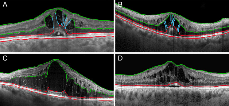
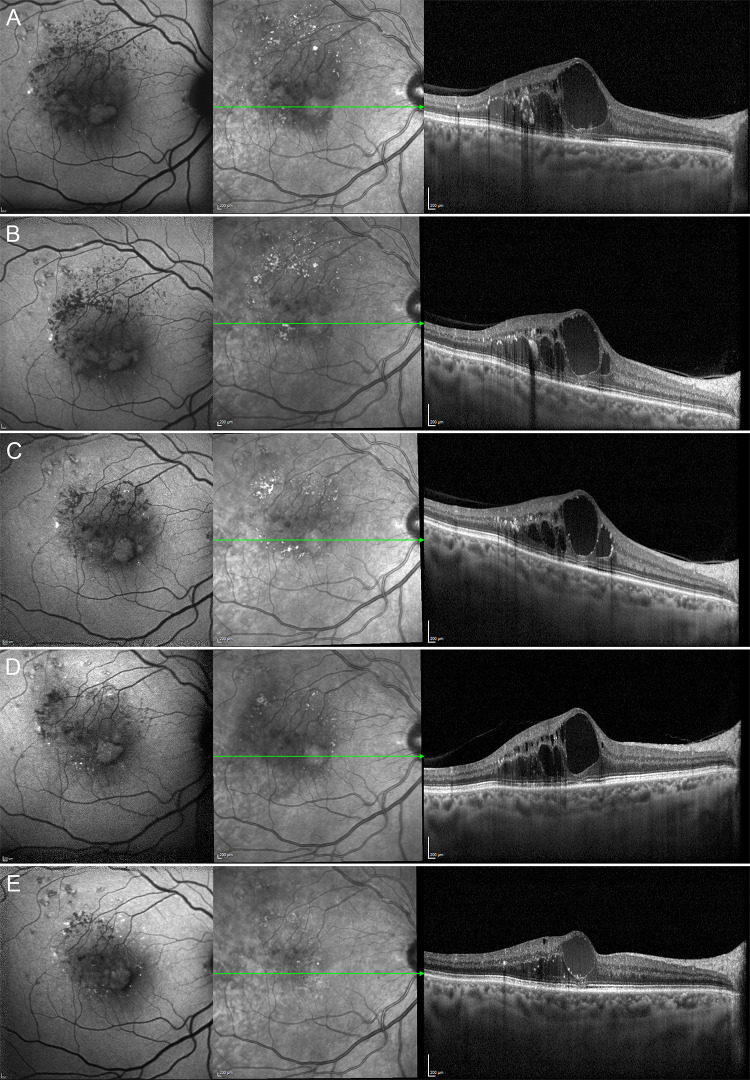
This study was a retrospective, observational case series. We included 168 eyes (168 patients) affected by central RVO (CRVO) and 116 eyes (116 patients) affected by branch (RVO). We collected clinical and imaging data from CRVO and BRVO eyes affected by macular edema with a minimum follow-up of 12 months. On structural OCT, we classified FE as pattern 1a, characterized by thick vertical intraretinal columns, pattern 1b, presenting thin vertical intraretinal lines, and pattern 2, showing no signs of vertical lines in the context of the cystoid macular edema. For statistical purposes, we considered data collected at baseline, after 1 year and at the last follow-up.

RESULTS

The mean follow-up was 40 ± 25 months for CRVO eyes and 36 ± 24 months for BRVO eyes. We found FE in 64 of 168 CRVO eyes (38%) and in 25 of 116 BRVO eyes (22%). Most of the eyes developed FE during the follow-up. For CRVO eyes, we found 6 eyes (9%) with pattern 1a, 17 eyes (26%) with pattern 1b and 41 eyes (65%) with pattern 2. Of those BRVO eyes with FE, we found 8 eyes (32%) with pattern 1a + 1b and 17 eyes (68%) with pattern 2. In both CRVO and BRVO the presence of FE was significantly associated with higher persistence of macular edema and worse outcome, with FE pattern 2 representing the most severe condition. Remarkably, FE patterns 1a and 1b were characterized by BCVA stability over the follow-up, whereas FE pattern 2 showed significant bestcorrected visual acuity (BCVA) worsening at the end of the follow-up.

CONCLUSIONS

FE can be considered a negative prognostic biomarker in RVO, associated with higher persistence of macular edema and worse visual outcome. Müller cell impairment might represent the pathogenic mechanism leading to the loss of macular structural support and impairment of fluid homeostasis.

摘要

引言

黄斑反转(FE)是最近通过光学相干断层扫描(OCT)发现的一种与糖尿病性黄斑水肿不良预后相关的现象。本研究的主要目的是探讨FE指标在视网膜静脉阻塞(RVO)诊断检查中的作用。

方法

本研究为回顾性观察病例系列。我们纳入了168例中心性视网膜静脉阻塞(CRVO)患者的168只眼和116例分支视网膜静脉阻塞(BRVO)患者的116只眼。我们收集了CRVO和BRVO合并黄斑水肿患者的临床和影像学数据,随访时间至少为12个月。在结构性OCT上,我们将FE分为1a型,其特征为视网膜内垂直柱状增厚;1b型,表现为视网膜内垂直细线;2型,在黄斑囊样水肿背景下无垂直线条迹象。为了进行统计学分析,我们考虑了基线、1年后和最后一次随访时收集的数据。

结果

CRVO组患者的平均随访时间为40±25个月,BRVO组为36±24个月。我们在168只CRVO眼中的64只(38%)和116只BRVO眼中的25只(22%)发现了FE。大多数眼睛在随访期间出现了FE。在CRVO眼中,我们发现6只(9%)为1a型,17只(26%)为1b型,41只(65%)为2型。在有FE的BRVO眼中,我们发现8只(32%)为1a + 1b型,17只(68%)为2型。在CRVO和BRVO中,FE的存在均与黄斑水肿的持续时间延长和预后较差显著相关,其中2型FE代表最严重的情况。值得注意的是,1a型和1b型FE的特征是随访期间最佳矫正视力(BCVA)稳定,而2型FE在随访结束时显示BCVA显著恶化。

结论

FE可被视为RVO的不良预后生物标志物,与黄斑水肿的持续时间延长和视力预后较差相关。Müller细胞损伤可能是导致黄斑结构支持丧失和液体稳态受损的致病机制。

https://cdn.ncbi.nlm.nih.gov/pmc/blobs/7475/10287597/27699f6d2bf8/40123_2023_734_Fig5_HTML.jpg
https://cdn.ncbi.nlm.nih.gov/pmc/blobs/7475/10287597/7837b891f75f/40123_2023_734_Fig1_HTML.jpg
https://cdn.ncbi.nlm.nih.gov/pmc/blobs/7475/10287597/c2eeeace16ee/40123_2023_734_Fig2_HTML.jpg
https://cdn.ncbi.nlm.nih.gov/pmc/blobs/7475/10287597/d24f88b81c74/40123_2023_734_Fig3_HTML.jpg
https://cdn.ncbi.nlm.nih.gov/pmc/blobs/7475/10287597/eda17f4960bb/40123_2023_734_Fig4_HTML.jpg
https://cdn.ncbi.nlm.nih.gov/pmc/blobs/7475/10287597/27699f6d2bf8/40123_2023_734_Fig5_HTML.jpg

相似文献

1
Foveal Eversion is Associated with High Persistence of Macular Edema and Visual Acuity Deterioration in Retinal Vein Occlusion.黄斑反转与视网膜静脉阻塞时黄斑水肿的高持续性及视力恶化相关。
Ophthalmol Ther. 2023 Aug;12(4):2157-2169. doi: 10.1007/s40123-023-00734-9. Epub 2023 Jun 9.
2
Longitudinal Assessment of the Choroidal Vascularity Index in Eyes with Branch Retinal Vein Occlusion-Associated Cystoid Macular Edema.视网膜分支静脉阻塞相关性黄斑囊样水肿患者脉络膜血管指数的纵向评估
Ophthalmol Ther. 2023 Aug;12(4):2103-2115. doi: 10.1007/s40123-023-00731-y. Epub 2023 May 23.
3
Collateral Vessel Development in Central and Branch Retinal Vein Occlusions Are Associated With Worse Visual and Anatomic Outcomes.中心性和分支性视网膜静脉阻塞的侧支血管发育与更差的视力和解剖学结局相关。
Invest Ophthalmol Vis Sci. 2021 Nov 1;62(14):1. doi: 10.1167/iovs.62.14.1.
4
[Analysis of visual prognosis and correlative factors in retinal vein occlusion].视网膜静脉阻塞的视觉预后及相关因素分析
Zhonghua Yan Ke Za Zhi. 2002 Feb;38(2):98-102.
5
Bevacizumab treatment of macular edema in CRVO and BRVO: long-term follow-up. (BERVOLT study: Bevacizumab for RVO long-term follow-up).贝伐单抗治疗视网膜中央静脉阻塞(CRVO)和视网膜分支静脉阻塞(BRVO)所致黄斑水肿:长期随访。(BERVOLT研究:贝伐单抗治疗视网膜静脉阻塞的长期随访)
Graefes Arch Clin Exp Ophthalmol. 2016 May;254(5):835-44. doi: 10.1007/s00417-015-3130-z. Epub 2015 Aug 14.
6
Grid photocoagulation combined with intravitreal bevacizumab for recurrent macular edema associated with retinal vein occlusion.格栅光凝联合玻璃体内注射贝伐单抗治疗视网膜静脉阻塞相关复发性黄斑水肿
Clin Ophthalmol. 2011;5:1031-6. doi: 10.2147/OPTH.S22923. Epub 2011 Jul 26.
7
Two or more dexamethasone intravitreal implants in treatment-naïve patients with macular edema due to retinal vein occlusion: subgroup analysis of a retrospective chart review study.在未经治疗的视网膜静脉阻塞所致黄斑水肿患者中使用两个或更多地塞米松玻璃体内植入物:一项回顾性病历审查研究的亚组分析
BMC Ophthalmol. 2015 Sep 4;15:118. doi: 10.1186/s12886-015-0106-z.
8
Intravitreal triamcinolone for the treatment of macular edema associated with central retinal vein occlusion.玻璃体内注射曲安奈德治疗视网膜中央静脉阻塞相关的黄斑水肿。
Arch Ophthalmol. 2004 Aug;122(8):1131-6. doi: 10.1001/archopht.122.8.1131.
9
Baseline predictors of visual acuity and retinal thickness outcomes in patients with retinal vein occlusion: Standard Care Versus COrticosteroid for REtinal Vein Occlusion Study report 10.视网膜静脉阻塞患者视力和视网膜厚度结果的基线预测因素:视网膜静脉阻塞标准护理与皮质类固醇治疗研究报告10
Ophthalmology. 2011 Feb;118(2):345-52. doi: 10.1016/j.ophtha.2010.06.034.
10
Vitrectomy for macular edema due to retinal vein occlusion.视网膜静脉阻塞所致黄斑水肿的玻璃体切除术
Clin Ophthalmol. 2019 Jun 13;13:969-984. doi: 10.2147/OPTH.S203212. eCollection 2019.

引用本文的文献

1
Global trends in retinal vein occlusion studies from 2004 to 2023: a bibliometric analysis.2004年至2023年视网膜静脉阻塞研究的全球趋势:一项文献计量分析
Int J Ophthalmol. 2025 Sep 18;18(9):1759-1769. doi: 10.18240/ijo.2025.09.18. eCollection 2025.
2
What is Occluding Our Understanding of Retinal Vein Occlusion?是什么阻碍了我们对视网膜静脉阻塞的理解?
Ophthalmol Ther. 2024 Dec;13(12):3025-3034. doi: 10.1007/s40123-024-01042-6. Epub 2024 Oct 10.
3
Digital histology of retinal microaneurysms as provided by dense B-scan (DART) OCTA: characteristics and clinical relevance in diabetic retinopathy.

本文引用的文献

1
Quantitative approaches in multimodal fundus imaging: State of the art and future perspectives.多模态眼底成像中的定量方法:现状与未来展望。
Prog Retin Eye Res. 2023 Jan;92:101111. doi: 10.1016/j.preteyeres.2022.101111. Epub 2022 Aug 4.
2
Foveal eversion patterns in diabetic macular edema.糖尿病性黄斑水肿的中心凹翻转模式。
Sci Rep. 2022 Jul 30;12(1):13097. doi: 10.1038/s41598-022-17555-8.
3
Retinal vein occlusion: drug targets and therapeutic implications.视网膜静脉阻塞:药物靶点和治疗意义。
DART OCTA 提供的视网膜微动脉瘤数字组织学:糖尿病视网膜病变中的特征和临床相关性。
Eye (Lond). 2024 Nov;38(16):3108-3112. doi: 10.1038/s41433-024-03230-x. Epub 2024 Jul 15.
4
Proteome Analysis of Bevacizumab Intervention in Experimental Central Retinal Vein Occlusion.贝伐单抗干预实验性视网膜中央静脉阻塞的蛋白质组学分析
J Pers Med. 2023 Nov 7;13(11):1580. doi: 10.3390/jpm13111580.
Expert Opin Ther Targets. 2021 Oct;25(10):847-864. doi: 10.1080/14728222.2021.2005026. Epub 2021 Dec 7.
4
Photocoagulation for retinal vein occlusion.视网膜静脉阻塞的光凝治疗
Prog Retin Eye Res. 2021 Nov;85:100964. doi: 10.1016/j.preteyeres.2021.100964. Epub 2021 Mar 11.
5
Real-Life Management of Central and Branch Retinal Vein Occlusion: A Seven-Year Follow-Up Study.真实世界中视网膜中央静脉和分支静脉阻塞的管理:一项为期七年的随访研究。
Thromb Haemost. 2021 Oct;121(10):1361-1366. doi: 10.1055/s-0041-1725197. Epub 2021 Mar 11.
6
Foveal Eversion: A Possible Biomarker of Persistent Diabetic Macular Edema.黄斑反转:持续性糖尿病性黄斑水肿的一种可能生物标志物。
Ophthalmol Ther. 2021 Mar;10(1):115-126. doi: 10.1007/s40123-020-00324-z. Epub 2021 Jan 9.
7
Glia of the human retina.人视网膜胶质细胞。
Glia. 2020 Apr;68(4):768-796. doi: 10.1002/glia.23727. Epub 2019 Dec 3.
8
Guidelines for the Management of Retinal Vein Occlusion by the European Society of Retina Specialists (EURETINA).欧洲视网膜专家学会(EURETINA)关于视网膜静脉阻塞管理的指南。
Ophthalmologica. 2019;242(3):123-162. doi: 10.1159/000502041. Epub 2019 Aug 14.
9
Ischemic retinal vein occlusion: characterizing the more severe spectrum of retinal vein occlusion.缺血性视网膜静脉阻塞:视网膜静脉阻塞更严重谱的特征。
Surv Ophthalmol. 2018 Nov-Dec;63(6):816-850. doi: 10.1016/j.survophthal.2018.04.005. Epub 2018 Apr 27.
10
Müller cells and diabetic retinopathy.米勒细胞与糖尿病视网膜病变
Vision Res. 2017 Oct;139:93-100. doi: 10.1016/j.visres.2017.03.013. Epub 2017 Sep 5.