De Benedictis Alessandro, Marasi Alessandra, Rossi-Espagnet Maria Camilla, Napolitano Antonio, Parrillo Chiara, Fracassi Donatella, Baldassari Giulia, Borro Luca, Bua Antonella, de Palma Luca, Luisi Concetta, Pepi Chiara, Savioli Alessandra, Luglietto Davide, Marras Carlo E
Neurosurgery Unit, Bambino Gesù Children's Hospital, IRCCS, 4, Piazza S. Onofrio, 00165 Rome, Italy.
Neuroradiology Unit, Bambino Gesù Children's Hospital, IRCCS, 4, Piazza S. Onofrio, 00165 Rome, Italy.
J Clin Med. 2023 May 31;12(11):3779. doi: 10.3390/jcm12113779.
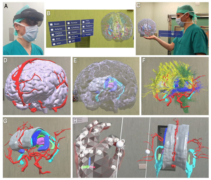
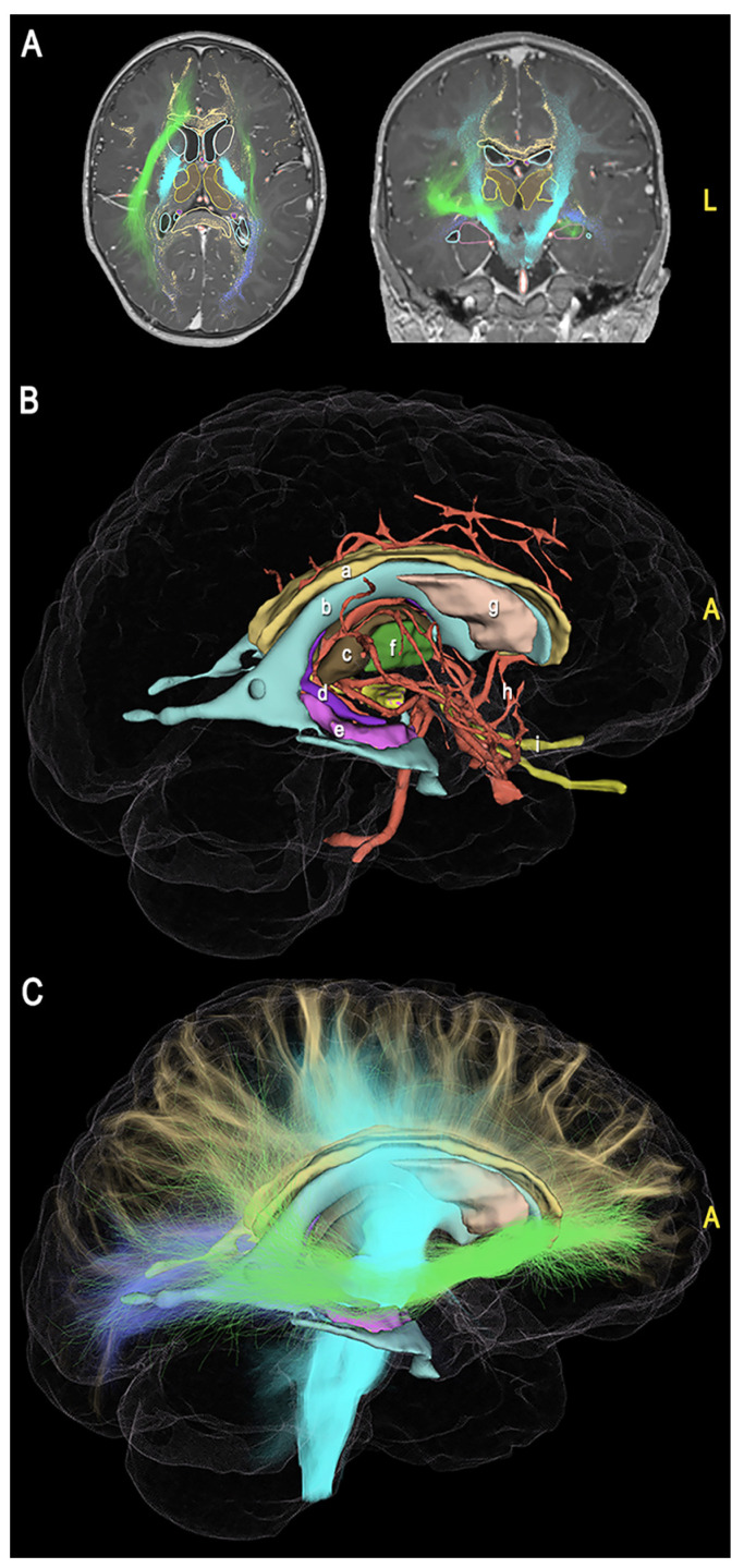
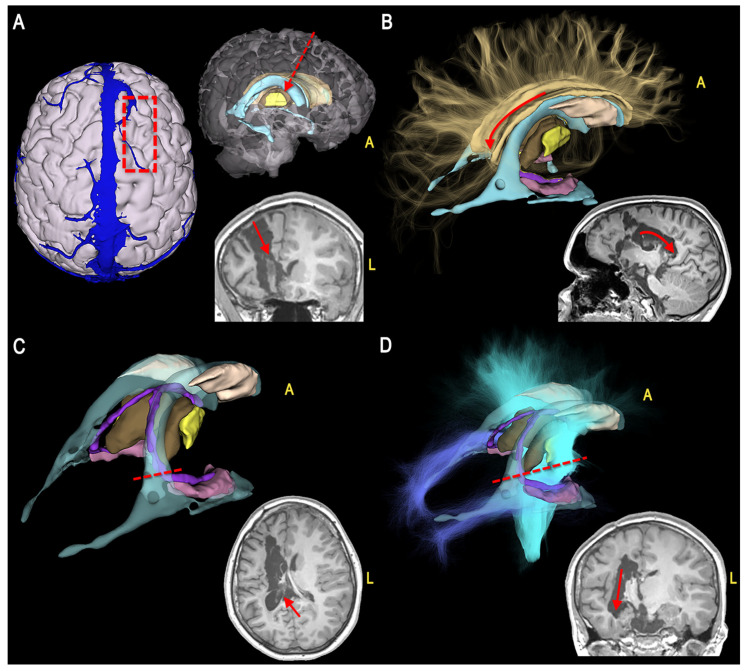
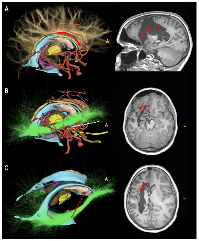
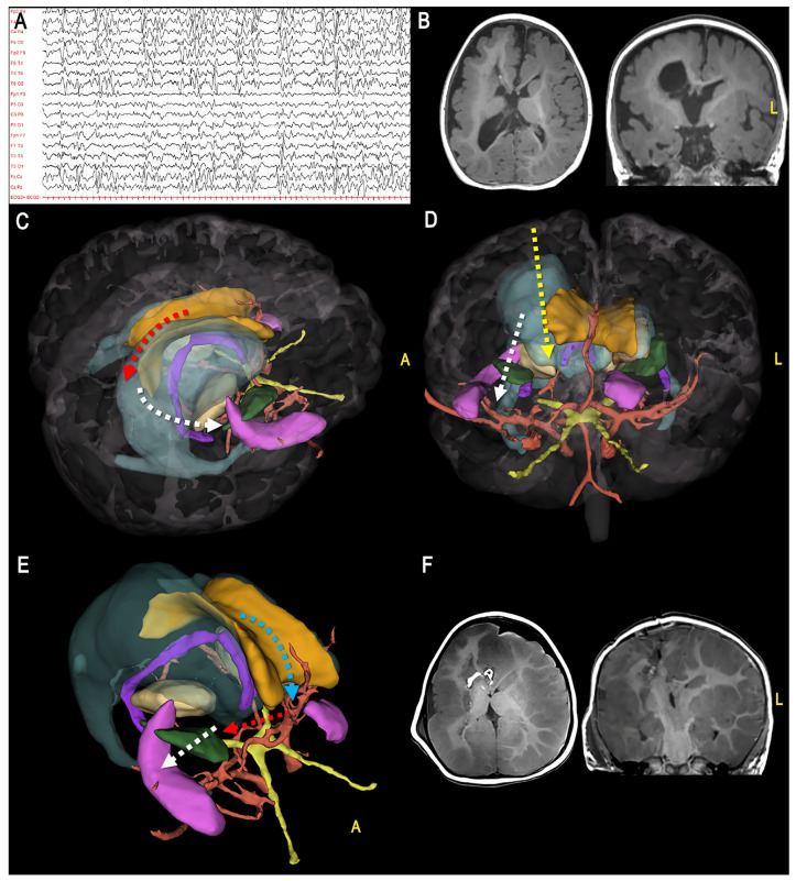
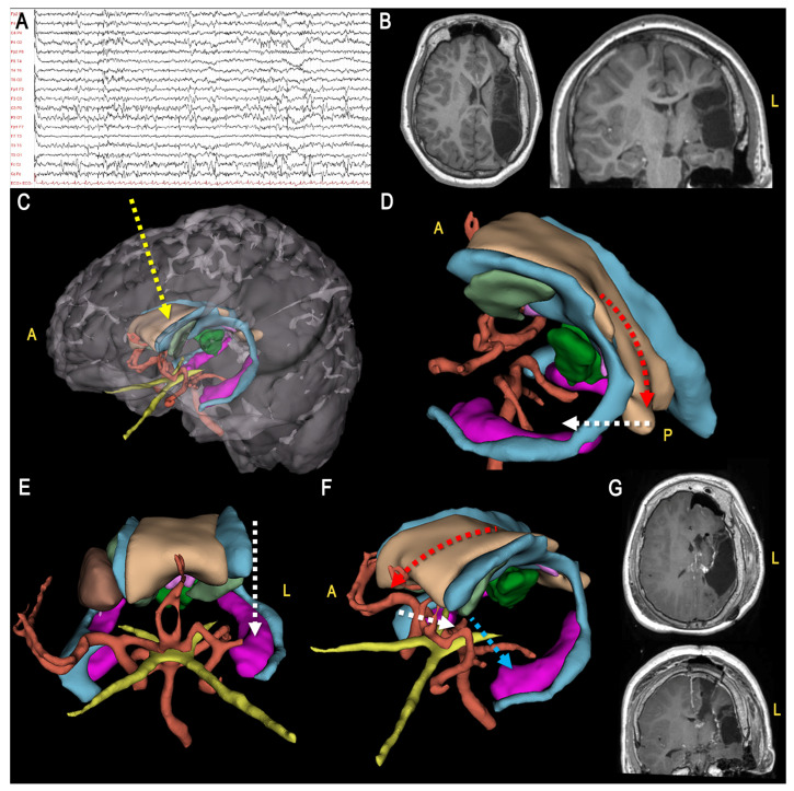
Vertical hemispherotomy is an effective treatment for many drug-resistant encephalopathies with unilateral involvement. One of the main factors influencing positive surgical results and long-term seizure freedom is the quality of disconnection. For this reason, perfect anatomical awareness is mandatory during each step of the procedure. Although previous groups attempted to reproduce the surgical anatomy through schematic representations, cadaveric dissections, and intraoperative photographs and videos, a comprehensive understanding of the approach may still be difficult, especially for less experienced neurosurgeons. In this work, we reported the application of advanced technology for three-dimensional (3D) modeling and visualization of the main neurova-scular structures during vertical hemispherotomy procedures. In the first part of the study, we built a detailed 3D model of the main structures and landmarks involved during each disconnection phase. In the second part, we discussed the adjunctive value of augmented reality systems for the management of the most challenging etiologies, such as hemimegalencephaly and post-ischemic encephalopathy. We demonstrated the contribution of advanced 3D modeling and visualization to enhance the quality of anatomical representation and interaction between the operator and model according to a surgical perspective, optimizing the quality of presurgical planning, intraoperative orientation, and educational training.
垂直性大脑半球切开术是治疗许多单侧受累的耐药性脑病的有效方法。影响手术阳性结果和长期无癫痫发作的主要因素之一是离断的质量。因此,在手术的每一步都必须具备完善的解剖学认知。尽管之前的研究团队试图通过示意图、尸体解剖以及术中照片和视频来重现手术解剖结构,但对该手术入路的全面理解可能仍然困难,尤其是对于经验不足的神经外科医生而言。在本研究中,我们报告了先进技术在垂直性大脑半球切开术过程中对主要神经血管结构进行三维(3D)建模和可视化的应用。在研究的第一部分,我们构建了每个离断阶段所涉及的主要结构和标志的详细3D模型。在第二部分,我们讨论了增强现实系统对于处理诸如半侧巨脑症和缺血性脑病等最具挑战性病因的辅助价值。我们展示了先进的3D建模和可视化技术在根据手术视角提高解剖结构呈现质量以及操作者与模型之间的交互方面的作用,优化了术前规划、术中定位和教育培训的质量。