Liu Gabrielle Y, Colangelo Laura A, Ash Samuel Y, San Jose Estepar Raul, Jacobs David R, Thyagarajan Bharat, Wells J Michael, Putman Rachel K, Choi Bina, Stevenson Christopher S, Carnethon Mercedes, Washko George R, Kalhan Ravi
Division of Pulmonary and Critical Care Medicine, Northwestern University Feinberg School of Medicine, Chicago, IL, USA.
Department of Preventive Medicine, Northwestern University Feinberg School of Medicine, Chicago, IL, USA.
ERJ Open Res. 2023 May 22;9(3). doi: 10.1183/23120541.00004-2023. eCollection 2023 May.
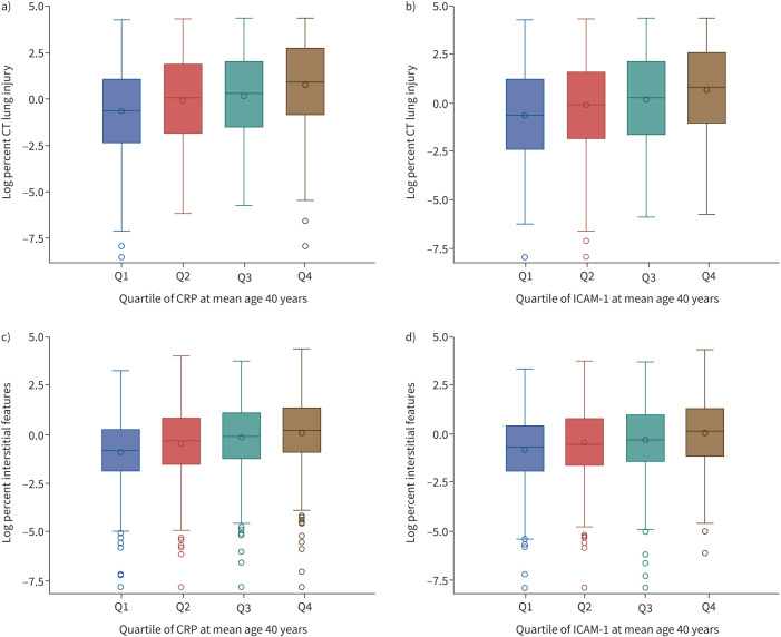
Visually normal areas of the lung with high attenuation on computed tomography (CT) imaging, termed CT lung injury, may represent injured but not yet remodelled lung parenchyma. This prospective cohort study examined if CT lung injury is associated with future interstitial features on CT and restrictive spirometry abnormality among participants from the Coronary Artery Risk Development in Young Adults (CARDIA) study.
CARDIA is a population-based cohort study. CT scans obtained at two time points were assessed objectively for amount of lung tissue characterised as CT lung injury and interstitial features. Restrictive spirometry was defined as having a forced vital capacity (FVC) <80% predicted with forced expiratory volume in 1 s/FVC ratio >70%.
Among 2213 participants, the median percentage of lung tissue characterised as CT lung injury at a mean age of 40 years was 3.4% (interquartile range 0.8-18.0%). After adjustment for covariates, a 10% higher amount of CT lung injury at mean age 40 years was associated with a 4.37% (95% CI 3.99-4.74%) higher amount of lung tissue characterised as interstitial features at mean age 50 years. Compared to those with the lowest quartile of CT lung injury at mean age 40 years, there were higher odds of incident restrictive spirometry at mean age 55 years in quartile 2 (OR 2.05, 95% CI 1.20-3.48), quartile 3 (OR 2.80, 95% CI 1.66-4.72) and quartile 4 (OR 3.77, 95% CI 2.24-6.33).
CT lung injury is an early objective measure that indicates risk of future lung impairment.
在计算机断层扫描(CT)成像上表现为高衰减的肺部视觉正常区域,称为CT肺损伤,可能代表受损但尚未重塑的肺实质。这项前瞻性队列研究调查了在年轻成人冠状动脉风险发展(CARDIA)研究的参与者中,CT肺损伤是否与未来CT上的间质特征及限制性肺量计异常相关。
CARDIA是一项基于人群的队列研究。对在两个时间点获得的CT扫描进行客观评估,以确定被表征为CT肺损伤和间质特征的肺组织量。限制性肺量计定义为用力肺活量(FVC)<预测值的80%且1秒用力呼气量/FVC比值>70%。
在2213名参与者中,平均年龄40岁时被表征为CT肺损伤的肺组织中位百分比为3.4%(四分位间距0.8 - 18.0%)。在调整协变量后,平均年龄40岁时CT肺损伤量每增加10%,与平均年龄50岁时被表征为间质特征的肺组织量增加4.37%(95%置信区间3.99 - 4.74%)相关。与平均年龄40岁时CT肺损伤处于最低四分位数的人相比,在平均年龄55岁时,四分位数2(比值比2.05,95%置信区间1.20 - 3.48)、四分位数3(比值比2.80,95%置信区间1.66 - 4.72)和四分位数4(比值比3.77,95%置信区间2.24 - 6.33)发生限制性肺量计异常的几率更高。
CT肺损伤是一种早期客观指标,表明未来肺部受损的风险。