Zwang Theodore J, Woost Benjamin, Bailey Joshua, Hoglund Zachary, Richardson Douglas S, Bennett Rachel E, Hyman Bradley T
Department of Neurology, MassGeneral Institute for Neurodegenerative Disease, Massachusetts General Hospital, Charlestown, MA, USA.
Harvard Medical School, Boston, MA, USA.
Brain Commun. 2023 Apr 19;5(3):fcad130. doi: 10.1093/braincomms/fcad130. eCollection 2023.
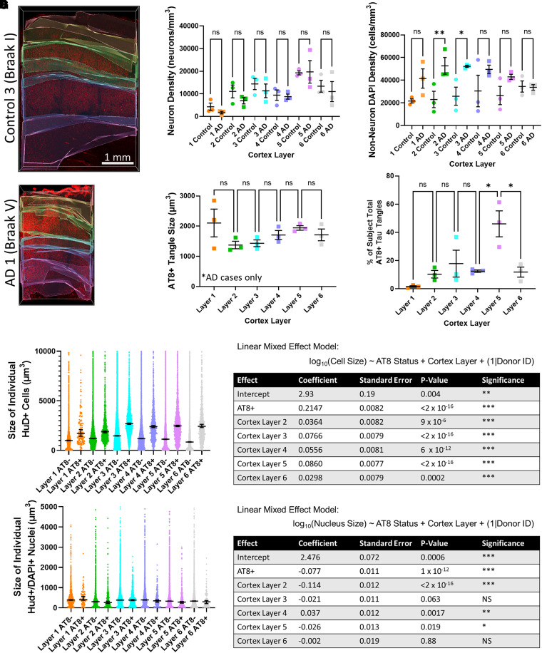
Studies of post-mortem human tissue provide insight into pathological processes, but are inherently limited by practical considerations that limit the scale at which tissue can be examined, and the obvious issue that the tissue reflects only one time point in a continuous disease process. We approached this problem by adapting new tissue clearance techniques to an entire cortical area of human brain, which allows surveillance of hundreds of thousands of neurons throughout the depth of the entire cortical thickness. This approach allows detection of 'rare' events that may be difficult to detect in standard 5 micrometre-thick paraffin sections. For example, it is well established that neurofibrillary tangles begin within a neuron, and ultimately, in at least some instances, persist in the brain even after the neuron has died. These are referred to as 'ghost tangles', a term that appropriately implies their 'difficult to see' ephemeral qualities. We set out to find ghost tangles as one example of the power of the tissue clearance/image analysis techniques to detect rare events, and to learn what happens at the end-point of a tangle's life history. We were able to identify 8103 tau tangles, 132 465 neurons and 299 640 nuclei in tissue samples from three subjects with severe Alzheimer's disease (Braak V-VI) and 4 tau tangles, 200 447 neurons and 462 715 nuclei in tissue samples from three subjects with no significant tau pathology (Braak 0-I). Among these data, we located 57 ghost tangles, which makes them only 0.7% of the total tau tangles observed. We found that ghost tangles are more likely to be found in cortical layers 3 and 5 (49/57), with a select few scattered across other layers 1, 2, 4 and 6. This ability to find rare events, such as ghost tangles, in large enough quantities to statistically test their distribution exemplifies how tissue clearing can be used as a powerful tool for studying selective vulnerability or resilience to pathology across brain regions.
对人体尸检组织的研究有助于深入了解病理过程,但由于实际因素的限制,其存在固有局限性,这些因素限制了可检查组织的规模,以及组织仅反映连续疾病过程中一个时间点这一明显问题。我们通过将新的组织清除技术应用于人类大脑的整个皮质区域来解决这个问题,这使得能够在整个皮质厚度的深度范围内监测数十万神经元。这种方法能够检测到在标准的5微米厚石蜡切片中可能难以检测到的“罕见”事件。例如,众所周知,神经原纤维缠结始于神经元内部,并且最终,至少在某些情况下,即使在神经元死亡后仍会在大脑中持续存在。这些被称为“幽灵缠结”,这个术语恰当地暗示了它们“难以看见”的短暂特性。我们着手寻找幽灵缠结,以此作为组织清除/图像分析技术检测罕见事件能力的一个例子,并了解缠结生命历程终点时会发生什么。我们能够在三名患有严重阿尔茨海默病(Braak V-VI)的受试者的组织样本中识别出8103个tau缠结、132465个神经元和299640个细胞核,在三名没有明显tau病理(Braak 0-I)的受试者的组织样本中识别出4个tau缠结、200447个神经元和462715个细胞核。在这些数据中,我们找到了57个幽灵缠结,这使得它们仅占观察到的总tau缠结的0.7%。我们发现幽灵缠结更有可能出现在皮质第3层和第5层(49/57),只有少数分散在其他第1、2、4和6层。能够找到足够数量的罕见事件,如幽灵缠结,以便对其分布进行统计测试,这例证了组织清除如何能够作为一种强大的工具,用于研究大脑区域对病理的选择性易损性或恢复力。