Vieira Warren A, Raymond Michael, Kelley Kristina, Cherubino Matthew A, Sahin Hande, McCusker Catherine D
McCusker Lab, Biology Department, University of Massachusetts Boston, Boston, MA, United States.
Front Cell Dev Biol. 2023 Jun 2;11:1152510. doi: 10.3389/fcell.2023.1152510. eCollection 2023.
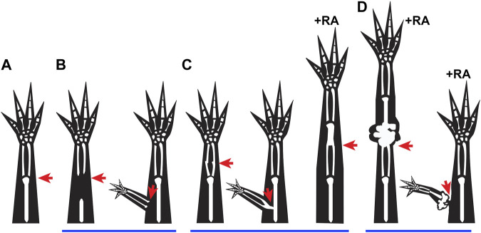
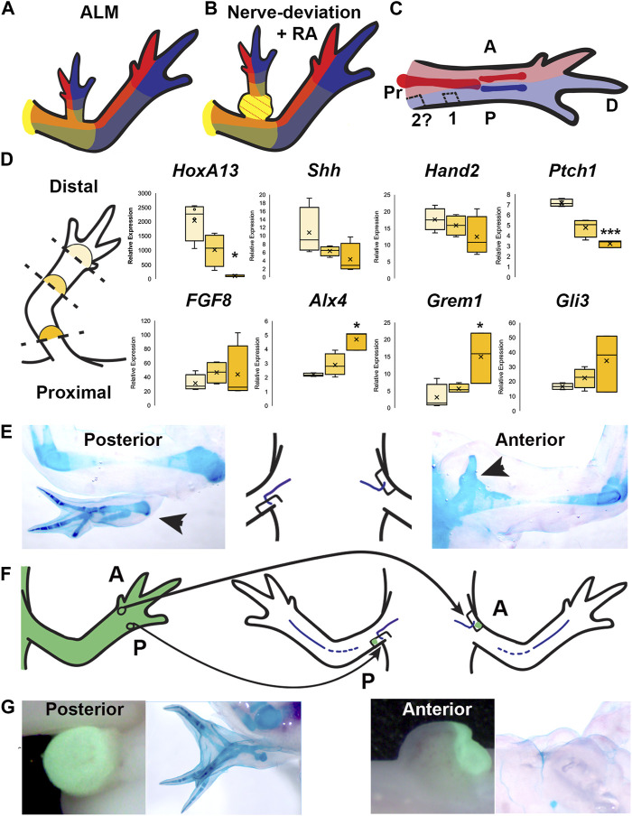
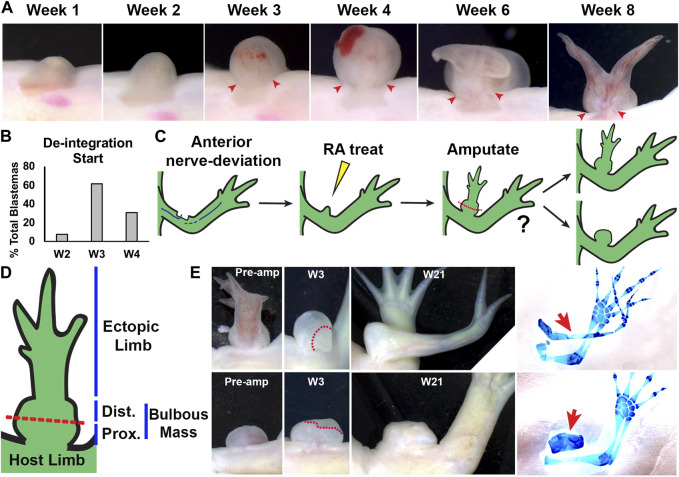
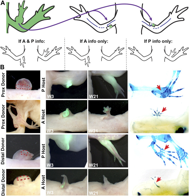
Little is known about how the newly regenerated limb tissues in the Mexican axolotl seamlessly integrate with the remaining stump tissues to form a functional structure, and why this doesn't occur in some regenerative scenarios. In this study, we evaluate the phenomenological and transcriptional characteristics associated with integration failure in ectopic limb structures generated by treating anterior-located ectopic blastemas with Retinoic Acid (RA) and focusing on the "bulbus mass" tissue that forms between the ectopic limb and the host site. We additionally test the hypothesis that the posterior portion of the limb base contains anterior positional identities. The positional identity of the bulbus mass was evaluated by assaying regenerative competency, the ability to induce new pattern in the Accessory Limb Model (ALM) assay, and by using qRTPCR to quantify the relative expression of patterning genes as the bulbus mass deintegrates from the host site. We additionally use the ALM and qRTPCR to analyze the distribution of anterior and posterior positional identities along the proximal/distal limb axis of uninjured and regenerating limbs. The bulbus mass regenerates limb structures with decreased complexity when amputated and is able to induce complex ectopic limb structure only when grafted into posterior-located ALMs. Expressional analysis shows significant differences in , , , , , and expression between the bulbus mass and the host site when deintegration is occuring. Grafts of posterior skin from the distal limb regions into posterior ALMs at the base of the limb induce ectopic limb structures. Proximally-located blastemas express significantly less and , and significantly more and than distally located blastemas. These findings show that the bulbus mass has an anterior-limb identity and that the expression of limb patterning genes is mismatched between the bulbus mass and the host limb. Our findings additionally show that anterior positional information is more abundant at the limb base, and that anterior patterning genes are more abundantly expressed in proximally located blastemas compared to blastemas in the more distal regions of the limb. These experiments provide valuable insight into the underlying causes of integration failure and further map the distribution of positional identities in the mature limb.
关于墨西哥钝口螈新再生的肢体组织如何与剩余的残端组织无缝整合以形成功能结构,以及为什么在某些再生情况下不会发生这种整合,我们知之甚少。在本研究中,我们评估了用视黄酸(RA)处理位于前部的异位芽基所产生的异位肢体结构中与整合失败相关的现象学和转录特征,并聚焦于在异位肢体与宿主部位之间形成的“球根状肿块”组织。我们还检验了肢体基部后部包含前部位置身份的假说。通过测定再生能力、在附属肢体模型(ALM)试验中诱导新模式的能力,以及在球根状肿块从宿主部位分离时使用qRTPCR定量模式形成基因的相对表达,来评估球根状肿块的位置身份。我们还使用ALM和qRTPCR分析未受伤和再生肢体沿近端/远端肢体轴的前部和后部位置身份的分布。球根状肿块在截肢后再生的肢体结构复杂性降低,并且仅当移植到位于后部的ALM中时才能诱导复杂的异位肢体结构。表达分析表明,在分离发生时,球根状肿块与宿主部位之间的、、、、和表达存在显著差异。将来自肢体远端区域的后部皮肤移植到肢体基部的后部ALM中可诱导异位肢体结构。与位于远端的芽基相比,位于近端的芽基表达的和显著更少,而和显著更多。这些发现表明,球根状肿块具有前部肢体身份,并且肢体模式形成基因在球根状肿块与宿主肢体之间的表达不匹配。我们的发现还表明,前部位置信息在肢体基部更为丰富,并且与肢体更远端区域的芽基相比,前部模式形成基因在位于近端的芽基中表达更为丰富。这些实验为整合失败的潜在原因提供了有价值的见解,并进一步描绘了成熟肢体中位置身份的分布。