Rao Aravindaksha, Deenadayalan P, Deepak C, Dilipkumar Dhivya, Angrish Nidhi, Shetty Suhani S
Department of Orthodontics and Dentofacial Orthopaedics, SRMKDC and H, Chennai, Tamil Nadu, India.
Private Practitioner, Bangalore, Karnataka, India.
J Orthod Sci. 2023 Mar 18;12:7. doi: 10.4103/jos.jos_52_22. eCollection 2023.
The aim of this in-vitro study was to observe and analyze the various enamel surface changes that occur due to laser debonding of metal and ceramic brackets, done by means of Er, Cr:YSGG laser.
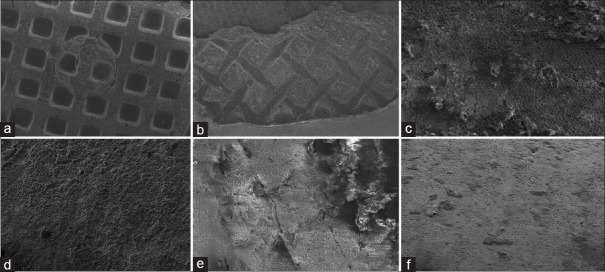
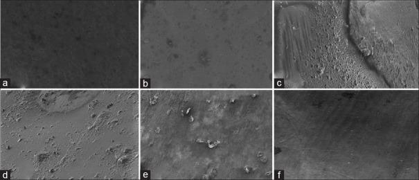
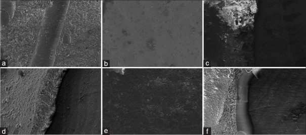
90 extracted premolars were randomly allocated into one of six groups with 15 teeth each. The groups represent metal brackets (Groups A1, A2, A3) and ceramic brackets (Groups B1, B2, B3). Each sub-group represents the mode of debonding used in the study. Debonded teeth were analyzed under scanning electron microscopy (SEM) at 80X and at 1000X magnification at three sites. The adhesive remnant index (ARI) scores were analyzed and the presence of enamel damage was observed.
ARI showed high score in Groups A1 and B1. SEM images of large composite remnants at the site of bracket in Groups A1 and B1 at the site of bracket and multiple enamel microcracks and fractures at interface and enamel adjacent to bracket in Groups A1 and B1. SEM images of minimal composite remnants at the site of bracket in Groups A2, A3, B2, and B3 and little to no presence of enamel microcracks or fractures at interface and enamel adjacent to bracket in Groups A2, A3, B2, and B3.
The use of Er, Cr:YSGG laser in orthodontic practice, especially in the debonding procedures of orthodontic brackets provide quality care to patient with minimal post-treatment damages.
本体外研究的目的是观察和分析通过铒铬:钇-钪-镓石榴石激光进行金属和陶瓷托槽激光脱粘时发生的各种牙釉质表面变化。
将90颗拔除的前磨牙随机分为6组,每组15颗牙。这些组代表金属托槽(A1、A2、A3组)和陶瓷托槽(B1、B2、B3组)。每个亚组代表研究中使用的脱粘方式。在扫描电子显微镜(SEM)下,以80倍和1000倍放大率在三个部位对脱粘后的牙齿进行分析。分析粘结剂残留指数(ARI)评分,并观察牙釉质损伤情况。
A1组和B1组的ARI得分较高。A1组和B1组托槽部位有大量复合树脂残留的SEM图像,A1组和B1组界面及托槽附近牙釉质有多处微裂纹和骨折。A2、A3、B2和B3组托槽部位复合树脂残留极少的SEM图像,A2、A3、B2和B3组界面及托槽附近牙釉质几乎没有微裂纹或骨折。
在正畸治疗中使用铒铬:钇-钪-镓石榴石激光,尤其是在正畸托槽的脱粘过程中,可为患者提供优质护理,使治疗后的损伤最小。