• 文献检索
  • 文档翻译
  • 深度研究
  • 学术资讯
  • Suppr Zotero 插件Zotero 插件
  • 邀请有礼
  • 套餐&价格
  • 历史记录
应用&插件
Suppr Zotero 插件Zotero 插件浏览器插件Mac 客户端Windows 客户端微信小程序
定价
高级版会员购买积分包购买API积分包
服务
文献检索文档翻译深度研究API 文档MCP 服务
关于我们
关于 Suppr公司介绍联系我们用户协议隐私条款
关注我们

Suppr 超能文献

核心技术专利:CN118964589B侵权必究
粤ICP备2023148730 号-1Suppr @ 2026

文献检索

告别复杂PubMed语法,用中文像聊天一样搜索,搜遍4000万医学文献。AI智能推荐,让科研检索更轻松。

立即免费搜索

文件翻译

保留排版,准确专业,支持PDF/Word/PPT等文件格式,支持 12+语言互译。

免费翻译文档

深度研究

AI帮你快速写综述,25分钟生成高质量综述,智能提取关键信息,辅助科研写作。

立即免费体验

一种带有 hRpn1 附着手柄的工程细胞系,用于分离蛋白酶体。

An engineered cell line with a hRpn1-attached handle to isolate proteasomes.

机构信息

Protein Processing Section, Structural Biophysics Laboratory, Center for Cancer Research, National Cancer Institute, National Institutes of Health, Frederick, Maryland, USA.

Genome Modification Core, Frederick National Laboratory for Cancer Research, Frederick, Maryland, USA.

出版信息

J Biol Chem. 2023 Aug;299(8):104948. doi: 10.1016/j.jbc.2023.104948. Epub 2023 Jun 22.

DOI:10.1016/j.jbc.2023.104948
PMID:37354974
原文链接:https://pmc.ncbi.nlm.nih.gov/articles/PMC10372910/
Abstract

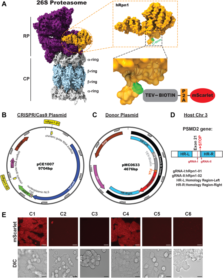
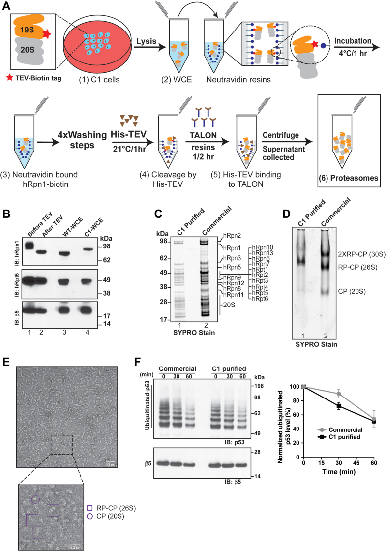
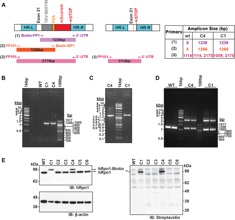
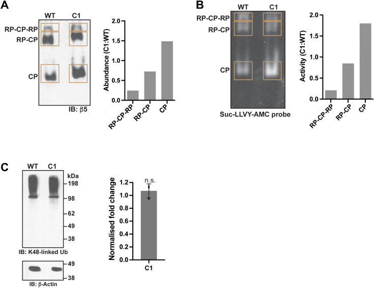
Regulated protein degradation in eukaryotes is performed by the 26S proteasome, which contains a 19-subunit regulatory particle (RP) that binds, processes, and translocates substrates to a 28-subunit hollow core particle (CP) where proteolysis occurs. In addition to its intrinsic subunits, myriad proteins interact with the proteasome transiently, including factors that assist and/or regulate its degradative activities. Efforts to identify proteasome-interacting components and/or to solve its structure have relied on over-expression of a tagged plasmid, establishing stable cell lines, or laborious purification protocols to isolate native proteasomes from cells. Here, we describe an engineered human cell line, derived from colon cancer HCT116 cells, with a biotin handle on the RP subunit hRpn1/PSMD2 (proteasome 26S subunit, non-ATPase 2) for purification of 26S proteasomes. A 75-residue sequence from Propionibacterium shermanii that is biotinylated in mammalian cells was added following a tobacco etch virus protease cut site at the C terminus of hRpn1. We tested and found that 26S proteasomes can be isolated from this modified HCT116 cell line by using a simple purification protocol. More specifically, biotinylated proteasomes were purified from the cell lysates by using neutravidin agarose resin and released from the resin following incubation with tobacco etch virus protease. The purified proteasomes had equivalent activity in degrading a model ubiquitinated substrate, namely ubiquitinated p53, compared to commercially available bovine proteasomes that were purified by fractionation. In conclusion, advantages of this approach to obtain 26S proteasomes over others is the simple purification protocol and that all cellular proteins, including the tagged hRpn1 subunit, remain at endogenous stoichiometry.

摘要

真核生物中的调节蛋白降解是由 26S 蛋白酶体完成的,该蛋白酶体包含一个由 19 个亚基组成的调节颗粒(RP),该颗粒结合、加工和转运底物到一个由 28 个亚基组成的空心核心颗粒(CP)中,在那里发生蛋白水解。除了其内在的亚基外,无数的蛋白质与蛋白酶体短暂相互作用,包括协助和/或调节其降解活性的因子。鉴定蛋白酶体相互作用成分和/或解决其结构的努力依赖于标记质粒的过表达、建立稳定的细胞系或费力的纯化方案,从细胞中分离天然蛋白酶体。在这里,我们描述了一种源自结肠癌 HCT116 细胞的工程化人细胞系,该细胞系在 RP 亚基 hRpn1/PSMD2(蛋白酶体 26S 亚基,非 ATP 酶 2)上带有生物素手柄,用于纯化 26S 蛋白酶体。烟草蚀纹病毒蛋白酶切割位点位于 hRpn1 的 C 末端后,在哺乳动物细胞中生物素化的短杆菌肽 S 来自 Propionibacterium shermanii 的 75 个残基序列。我们进行了测试,发现可以通过使用简单的纯化方案从该修饰的 HCT116 细胞系中分离 26S 蛋白酶体。更具体地说,通过使用链霉亲和素琼脂糖树脂从细胞裂解物中纯化生物素化的蛋白酶体,并在用烟草蚀纹病毒蛋白酶孵育后从树脂中释放。与通过分级分离纯化的商业上可获得的牛蛋白酶体相比,纯化的蛋白酶体在降解模型泛素化底物(即泛素化 p53)方面具有同等的活性。总之,与其他方法相比,这种获得 26S 蛋白酶体的方法的优点是简单的纯化方案以及所有细胞蛋白,包括标记的 hRpn1 亚基,都保持内源性比例。

https://cdn.ncbi.nlm.nih.gov/pmc/blobs/ec9b/10372910/379f0e9dc51c/gr5.jpg
https://cdn.ncbi.nlm.nih.gov/pmc/blobs/ec9b/10372910/76e1a501ab67/gr1.jpg
https://cdn.ncbi.nlm.nih.gov/pmc/blobs/ec9b/10372910/a89c0c2b9af9/gr2.jpg
https://cdn.ncbi.nlm.nih.gov/pmc/blobs/ec9b/10372910/cb7c5c3dafce/gr3.jpg
https://cdn.ncbi.nlm.nih.gov/pmc/blobs/ec9b/10372910/8656ecc18cd2/gr4.jpg
https://cdn.ncbi.nlm.nih.gov/pmc/blobs/ec9b/10372910/379f0e9dc51c/gr5.jpg

相似文献

1
An engineered cell line with a hRpn1-attached handle to isolate proteasomes.一种带有 hRpn1 附着手柄的工程细胞系,用于分离蛋白酶体。
J Biol Chem. 2023 Aug;299(8):104948. doi: 10.1016/j.jbc.2023.104948. Epub 2023 Jun 22.
2
Proteasomes: Isolation and Activity Assays.蛋白酶体:分离与活性测定。
Curr Protoc. 2023 Apr;3(4):e717. doi: 10.1002/cpz1.717.
3
The deubiquitinating enzyme Usp14 allosterically inhibits multiple proteasomal activities and ubiquitin-independent proteolysis.去泛素化酶Usp14通过变构作用抑制多种蛋白酶体活性和不依赖泛素的蛋白水解作用。
J Biol Chem. 2017 Jun 9;292(23):9830-9839. doi: 10.1074/jbc.M116.763128. Epub 2017 Apr 17.
4
Assembly chaperone Nas6 selectively destabilizes 26S proteasomes with defective regulatory particle-core particle interfaces.组装伴侣 Nas6 选择性地使具有缺陷的调节颗粒-核心颗粒界面的 26S 蛋白酶体不稳定。
J Biol Chem. 2023 Feb;299(2):102894. doi: 10.1016/j.jbc.2023.102894. Epub 2023 Jan 10.
5
Method of Monitoring 26S Proteasome in Cells Revealed the Crucial Role of PSMA3 C-Terminus in 26S Integrity.细胞中 26S 蛋白酶体监测方法揭示了 PSMA3 C 端在 26S 完整性中的关键作用。
Biomolecules. 2023 Jun 15;13(6):992. doi: 10.3390/biom13060992.
6
Mammalian 26S proteasomes remain intact during protein degradation.哺乳动物的26S蛋白酶体在蛋白质降解过程中保持完整。
Cell. 2008 Oct 17;135(2):355-65. doi: 10.1016/j.cell.2008.08.032.
7
26S proteasomes become stably activated upon heat shock when ubiquitination and protein degradation increase.26S 蛋白酶体在热休克时通过泛素化和蛋白质降解的增加而稳定激活。
Proc Natl Acad Sci U S A. 2022 Jun 21;119(25):e2122482119. doi: 10.1073/pnas.2122482119. Epub 2022 Jun 15.
8
NOXA, a sensor of proteasome integrity, is degraded by 26S proteasomes by an ubiquitin-independent pathway that is blocked by MCL-1.NOXA 是蛋白酶体完整性的传感器,它通过一种不依赖泛素的途径被 26S 蛋白酶体降解,该途径被 MCL-1 阻断。
Cell Death Differ. 2012 Sep;19(9):1424-34. doi: 10.1038/cdd.2012.16. Epub 2012 Feb 24.
9
cAMP-induced phosphorylation of 26S proteasomes on Rpn6/PSMD11 enhances their activity and the degradation of misfolded proteins.环磷酸腺苷(cAMP)诱导的26S蛋白酶体在Rpn6/PSMD11上的磷酸化增强了它们的活性以及错误折叠蛋白质的降解。
Proc Natl Acad Sci U S A. 2015 Dec 29;112(52):E7176-85. doi: 10.1073/pnas.1522332112. Epub 2015 Dec 15.
10
Measurement of the Multiple Activities of 26S Proteasomes.26S蛋白酶体多种活性的测定
Methods Mol Biol. 2018;1844:289-308. doi: 10.1007/978-1-4939-8706-1_19.

引用本文的文献

1
Structures of dynamic interactors at native proteasomes by PhIX-MS and cryoelectron microscopy.通过噬菌体展示-质谱联用技术和冷冻电子显微镜解析天然蛋白酶体中动态相互作用分子的结构。
bioRxiv. 2025 Aug 2:2025.07.31.667872. doi: 10.1101/2025.07.31.667872.
2
hRpn13 shapes the proteome and transcriptome through epigenetic factors HDAC8, PADI4, and transcription factor NF-κB p50.hRpn13 通过表观遗传因子 HDAC8、PADI4 和转录因子 NF-κB p50 塑造蛋白质组和转录组。
Mol Cell. 2024 Feb 1;84(3):522-537.e8. doi: 10.1016/j.molcel.2023.11.035. Epub 2023 Dec 26.