Department of Radiation Oncology, Ortenau Klinikum Offenburg-Kehl, Offenburg, Germany.
UKSH, Campus Kiel, Clinic of Anesthesiology and Operative Medicine, Kiel, Germany.
J Appl Clin Med Phys. 2023 Aug;24(8):e14077. doi: 10.1002/acm2.14077. Epub 2023 Jun 26.
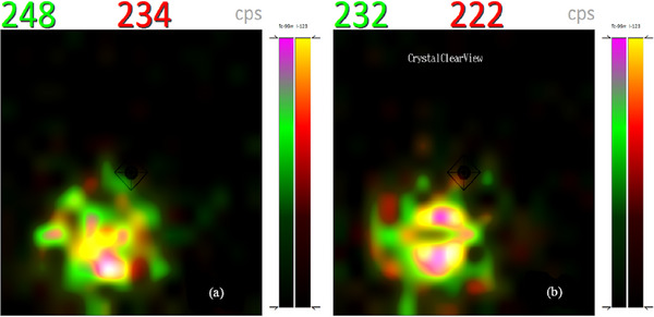
Performing lymphoscintigraphy in a separate room, frees up the conventional gamma camera, coupled with the desire to directly localize sentinel lymph nodes (SLN) in the operating theatre has led to the development of high-resolution semiconductor-detector based handheld gamma-cameras, CrystalCam.
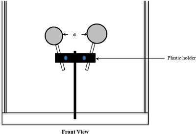
This work consists of phantom and clinical studies. For the first part, a Jaszczak phantom with hollow spheres of various volumes were filled with the Tc and the camera's sensitivity was measured at various distances to assess the possibilities and limitations of the device. The clinical study evaluates the effectiveness of CrystalCam in localizing SLN in 40 consecutive malignant melanoma patients compared to both conventional planar lymphoscintigraphy and hybrid SPECT/CT. SLNs detected by planar lymphoscintigraphy were marked on the patients' skin using a UV-marker. CrystalCam images were acquired in another room by another examiner and the SLNs were marked with a felt pen. The detected nodes by both camera systems were evaluated using UV-lamp and normal light to visualize the UV- and felt pen marks respectively. The concordance rate of the SLNs and higher-echelon nodes localized by both planar scintigraphy and CrystalCam imaging with respect to the total SLNs and higher-echelon nodes detected by SPECT/CT imaging are compared and statistically analyzed.
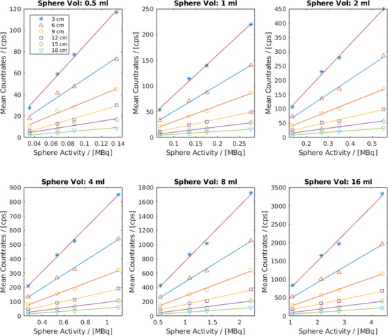
The results of the phantom study show a good correlation between activity and count-rates for all distancesSPECT/CT, CrystalCamm, and planar lymphoscintigraphy detected 69, 58, and 61 SLNs respectively. The concordance rate of 95.65% by the CrystalCam and planar scintigraphy implies both cameras are statistically coequal in preoperative SLN detection of malignant melanoma. For the higher-echelon nodes, SPECT/CT, planar and CrystalCam imaging systems identified 82, 48, and 13 respectively; thus, CrystalCam was statistically inferior to planar imaging.
The handheld CrystalCam is a reliable instrument for localizing SLNs in surgical centers without an on-site nuclear medicine department.
在单独的房间进行淋巴闪烁显像,可以释放传统的伽玛相机,并且希望直接在手术室定位前哨淋巴结 (SLN),这导致了基于高分辨率半导体探测器的手持式伽玛相机 CrystalCam 的发展。
这项工作包括体模和临床研究。对于第一部分,使用具有各种体积的空心球体的 Jaszczak 体模填充 Tc,并且在各种距离处测量相机的灵敏度,以评估该设备的可能性和局限性。临床研究评估了 CrystalCam 在定位 40 例连续恶性黑色素瘤患者的 SLN 方面的有效性,与传统的平面淋巴闪烁显像和混合 SPECT/CT 相比。使用 UV 标记器在患者皮肤上标记通过平面淋巴闪烁显像检测到的 SLN。由另一位检查人员在另一个房间中获取 CrystalCam 图像,并使用毡尖笔标记 SLN。使用 UV 灯和自然光分别可视化 UV 和毡尖笔标记,评估两个相机系统检测到的节点。使用 SPECT/CT 成像检测到的所有 SLN 和更高层次的节点,比较并统计分析平面闪烁显像和 CrystalCam 成像定位的 SLN 和更高层次的节点的符合率。
体模研究的结果显示,对于所有距离,活性和计数率之间都存在良好的相关性 SPECT/CT、CrystalCam 和平面淋巴闪烁显像分别检测到 69、58 和 61 个 SLN。CrystalCam 和平面闪烁显像的符合率为 95.65%,这意味着这两个相机在恶性黑色素瘤的术前 SLN 检测中在统计学上是相等的。对于更高层次的节点,SPECT/CT、平面和 CrystalCam 成像系统分别识别出 82、48 和 13 个;因此,CrystalCam 在统计学上不如平面成像。
手持式 CrystalCam 是在没有现场核医学部门的手术中心定位 SLN 的可靠仪器。