• 文献检索
  • 文档翻译
  • 深度研究
  • 学术资讯
  • Suppr Zotero 插件Zotero 插件
  • 邀请有礼
  • 套餐&价格
  • 历史记录
应用&插件
Suppr Zotero 插件Zotero 插件浏览器插件Mac 客户端Windows 客户端微信小程序
定价
高级版会员购买积分包购买API积分包
服务
文献检索文档翻译深度研究API 文档MCP 服务
关于我们
关于 Suppr公司介绍联系我们用户协议隐私条款
关注我们

Suppr 超能文献

核心技术专利:CN118964589B侵权必究
粤ICP备2023148730 号-1Suppr @ 2026

文献检索

告别复杂PubMed语法,用中文像聊天一样搜索,搜遍4000万医学文献。AI智能推荐,让科研检索更轻松。

立即免费搜索

文件翻译

保留排版,准确专业,支持PDF/Word/PPT等文件格式,支持 12+语言互译。

免费翻译文档

深度研究

AI帮你快速写综述,25分钟生成高质量综述,智能提取关键信息,辅助科研写作。

立即免费体验

利用 shotgun 蛋白质组学鉴定 Fuchs 内皮角膜营养不良患者的 Descemet 膜中表达的蛋白质。

Shotgun proteomics identification of proteins expressed in the Descemet's membrane of patients with Fuchs endothelial corneal dystrophy.

机构信息

Department of Biomedical Engineering, Faculty of Life and Medical Sciences, Doshisha University, Kyotanabe, 610-0394, Japan.

Genomics, Proteomics and Biomedical Functions, Graduate School of Life and Medical Sciences, Doshisha University, Kyoto, Japan.

出版信息

Sci Rep. 2023 Jun 27;13(1):10401. doi: 10.1038/s41598-023-37104-1.

DOI:10.1038/s41598-023-37104-1
PMID:37369713
原文链接:https://pmc.ncbi.nlm.nih.gov/articles/PMC10300001/
Abstract

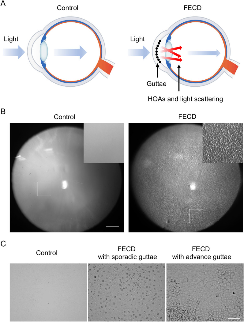
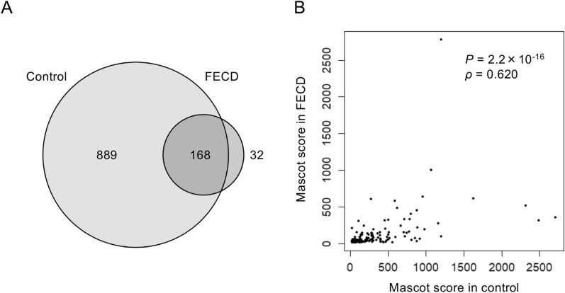
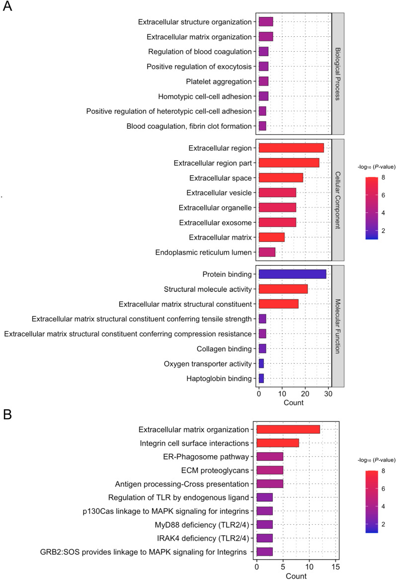
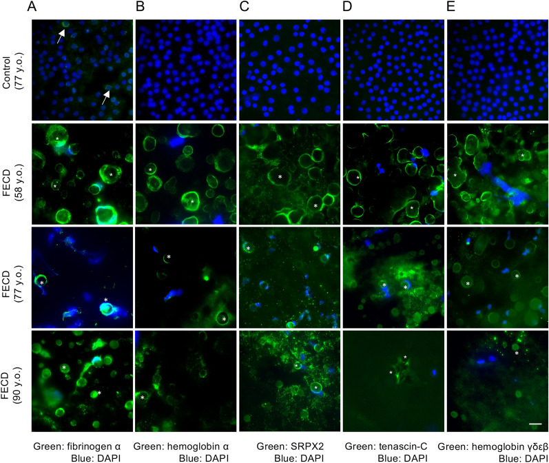
Fuchs endothelial corneal dystrophy (FECD) is a slowly evolving, bilateral disease of the corneal endothelium, characterized by an abnormal accumulation of extracellular matrix (ECM) in the basement membrane (Descemet's membrane, DM). This results in the formation of small round excrescences, called guttae, and a progressive disappearance of endothelial cells. In the intermediate stage, the numerous guttae create significant optical aberrations, and in the late stage, the loss of endothelial function leads to permanent corneal edema. The molecular components of guttae have not been fully elucidated. In the current study, we conducted shotgun proteomics of the DMs, including guttae, obtained from patients with FECD and revealed that 32 proteins were expressed only in the FECD-DMs but not in the DMs of control subjects. Subsequent enrichment analyses identified associations with multiple ECM-related pathways. Immunostaining of flat-mounted DMs confirmed that 4 of the top 5 identified proteins (hemoglobin α, SRPX2, tenascin-C, and hemoglobin γδεβ) were expressed in FECD-DMs but not in non-FECD-DMs. Fibrinogen α was strongly expressed in FECD-DMs, but weakly expressed in non-FECD-DMs. We also demonstrated that matrix-assisted laser desorption ionization imaging mass spectrometry (MALDI-IMS) can display the in situ spatial distribution of biomolecules expressed in the DM, including the guttae.

摘要

Fuchs 内皮角膜营养不良(FECD)是一种缓慢进展的、双侧角膜内皮疾病,其特征是细胞外基质(ECM)在基底膜(Descemet 膜,DM)中异常积聚。这导致形成小圆形赘生物,称为小滴,并伴有内皮细胞的逐渐消失。在中期,大量的小滴会产生明显的光学像差,而在晚期,内皮功能的丧失会导致永久性角膜水肿。小滴的分子成分尚未完全阐明。在本研究中,我们对包括小滴在内的来自 FECD 患者的 DM 进行了鸟枪法蛋白质组学分析,结果显示 32 种蛋白质仅在 FECD-DM 中表达,而不在对照组 DM 中表达。随后的富集分析确定了与多种 ECM 相关途径的关联。对平铺 DM 的免疫染色证实,在鉴定出的前 5 种蛋白(血红蛋白α、SRPX2、腱糖蛋白 C 和血红蛋白γδεβ)中,有 4 种(血红蛋白α、SRPX2、腱糖蛋白 C 和血红蛋白γδεβ)在 FECD-DM 中表达,而不在非 FECD-DM 中表达。纤维蛋白原α在 FECD-DM 中表达强烈,而非 FECD-DM 中表达较弱。我们还证明,基质辅助激光解吸电离成像质谱(MALDI-IMS)可以显示 DM 中表达的生物分子的原位空间分布,包括小滴。

https://cdn.ncbi.nlm.nih.gov/pmc/blobs/cd2e/10300001/b95b6e85c28e/41598_2023_37104_Fig5_HTML.jpg
https://cdn.ncbi.nlm.nih.gov/pmc/blobs/cd2e/10300001/893efef1f4ff/41598_2023_37104_Fig1_HTML.jpg
https://cdn.ncbi.nlm.nih.gov/pmc/blobs/cd2e/10300001/c6220ba76a1b/41598_2023_37104_Fig2_HTML.jpg
https://cdn.ncbi.nlm.nih.gov/pmc/blobs/cd2e/10300001/2aad26cef975/41598_2023_37104_Fig3_HTML.jpg
https://cdn.ncbi.nlm.nih.gov/pmc/blobs/cd2e/10300001/5864a763944d/41598_2023_37104_Fig4_HTML.jpg
https://cdn.ncbi.nlm.nih.gov/pmc/blobs/cd2e/10300001/b95b6e85c28e/41598_2023_37104_Fig5_HTML.jpg

相似文献

1
Shotgun proteomics identification of proteins expressed in the Descemet's membrane of patients with Fuchs endothelial corneal dystrophy.利用 shotgun 蛋白质组学鉴定 Fuchs 内皮角膜营养不良患者的 Descemet 膜中表达的蛋白质。
Sci Rep. 2023 Jun 27;13(1):10401. doi: 10.1038/s41598-023-37104-1.
2
Expression and Impact of Fibronectin, Tenascin-C, Osteopontin, and Type XIV Collagen in Fuchs Endothelial Corneal Dystrophy.纤连蛋白、肌腱蛋白-C、骨桥蛋白和 XIV 型胶原蛋白在富克斯内皮性角膜营养不良中的表达及影响
Invest Ophthalmol Vis Sci. 2024 Apr 1;65(4):38. doi: 10.1167/iovs.65.4.38.
3
Matrix metalloproteinases and their inhibitors in Fuchs endothelial corneal dystrophy.基质金属蛋白酶及其抑制剂在 Fuchs 内皮角膜营养不良中的作用。
Exp Eye Res. 2021 Apr;205:108500. doi: 10.1016/j.exer.2021.108500. Epub 2021 Feb 19.
4
Association of the Gutta-Induced Microenvironment With Corneal Endothelial Cell Behavior and Demise in Fuchs Endothelial Corneal Dystrophy.胶诱导微环境与 Fuchs 内皮角膜营养不良中角膜内皮细胞行为和死亡的关系。
JAMA Ophthalmol. 2018 Aug 1;136(8):886-892. doi: 10.1001/jamaophthalmol.2018.2031.
5
The soil and the seed: The relationship between Descemet's membrane and the corneal endothelium.土壤与种子:角膜后弹力层与角膜内皮细胞的关系。
Exp Eye Res. 2023 Feb;227:109376. doi: 10.1016/j.exer.2022.109376. Epub 2022 Dec 30.
6
Colocalization of increased transforming growth factor-beta-induced protein (TGFBIp) and Clusterin in Fuchs endothelial corneal dystrophy.在Fuchs内皮性角膜营养不良中,转化生长因子-β诱导蛋白(TGFBIp)增加与聚集蛋白的共定位。
Invest Ophthalmol Vis Sci. 2009 Mar;50(3):1129-36. doi: 10.1167/iovs.08-2525. Epub 2008 Nov 14.
7
Reduced expression of apolipoprotein E and immunoglobulin heavy constant gamma 1 proteins in Fuchs endothelial corneal dystrophy.载脂蛋白 E 和免疫球蛋白重链 γ1 蛋白在 Fuchs 内皮角膜营养不良中的表达降低。
Clin Exp Ophthalmol. 2019 Nov;47(8):1028-1042. doi: 10.1111/ceo.13569. Epub 2019 Jul 5.
8
Advanced glycation end products and receptors in Fuchs' dystrophy corneas undergoing Descemet's stripping with endothelial keratoplasty.在接受Descemet膜剥除联合内皮角膜移植术的Fuchs角膜内皮营养不良角膜中晚期糖基化终末产物及受体
Ophthalmology. 2007 Aug;114(8):1453-60. doi: 10.1016/j.ophtha.2006.10.049. Epub 2007 Feb 22.
9
Rapid detection of guttae area using aniline blue staining in Fuchs endothelial corneal dystrophy mouse model.使用苯胺蓝染色快速检测 Fuchs 内皮角膜营养不良小鼠模型中的角膜小滴区。
Clin Exp Pharmacol Physiol. 2024 Oct;51(10):e13921. doi: 10.1111/1440-1681.13921.
10
Biomechanical changes to Descemet's membrane precede endothelial cell loss in an early-onset murine model of Fuchs endothelial corneal dystrophy.生物力学变化对 Descemet 膜的影响早于内皮细胞丢失在一个早期发病的鼠模型的 Fuchs 内皮角膜营养不良。
Exp Eye Res. 2019 Mar;180:18-22. doi: 10.1016/j.exer.2018.11.021. Epub 2018 Nov 22.

引用本文的文献

1
A feasibility of computational drug screening for Fuchs endothelial corneal dystrophy.针对富克斯角膜内皮营养不良进行计算药物筛选的可行性。
Sci Rep. 2025 Apr 26;15(1):14665. doi: 10.1038/s41598-025-95003-z.
2
Comprehensive identification of dysregulated extracellular matrix molecules in the corneal endothelium of patients with Fuchs endothelial corneal dystrophy.全面鉴定Fuchs内皮性角膜营养不良患者角膜内皮中失调的细胞外基质分子。
Sci Rep. 2025 Apr 26;15(1):14654. doi: 10.1038/s41598-025-91850-y.
3
How "Omics" Studies Contribute to a Better Understanding of Fuchs' Endothelial Corneal Dystrophy.

本文引用的文献

1
RNA-Seq-based transcriptome analysis of corneal endothelial cells derived from patients with Fuchs endothelial corneal dystrophy.基于 RNA-Seq 的 Fuchs 内皮角膜营养不良患者角膜内皮细胞转录组分析。
Sci Rep. 2023 May 27;13(1):8647. doi: 10.1038/s41598-023-35468-y.
2
Recurrence of Guttae and Endothelial Dysfunction After Successful Descemet Stripping Only in Fuchs Dystrophy.仅在 Fuchs 营养不良中成功进行 Descemet 膜撕除术后出现胶滴和内皮功能障碍的复发。
Cornea. 2023 Aug 1;42(8):1037-1040. doi: 10.1097/ICO.0000000000003221. Epub 2022 Dec 19.
3
Applications of MALDI-MS/MS-Based Proteomics in Biomedical Research.
“组学”研究如何有助于更好地理解富克斯角膜内皮营养不良。
Curr Issues Mol Biol. 2025 Feb 20;47(3):135. doi: 10.3390/cimb47030135.
4
The TCF4 Gene Regulates Apoptosis of Corneal Endothelial Cells in Fuchs Endothelial Corneal Dystrophy.TCF4基因调控富克斯内皮性角膜营养不良中角膜内皮细胞的凋亡。
Invest Ophthalmol Vis Sci. 2025 Mar 3;66(3):16. doi: 10.1167/iovs.66.3.16.
5
Endoplasmic Reticulum-Mitochondria Crosstalk in Fuchs Endothelial Corneal Dystrophy: Current Status and Future Prospects.富克斯内皮性角膜营养不良中内质网与线粒体的相互作用:现状与未来展望
Int J Mol Sci. 2025 Jan 22;26(3):894. doi: 10.3390/ijms26030894.
6
Integrated Spatial Multi-Omics Study of Postmortem Brains of Alzheimer's Disease.阿尔茨海默病死后大脑的综合空间多组学研究
Acta Histochem Cytochem. 2024 Jun 28;57(3):119-130. doi: 10.1267/ahc.24-00025.
7
Senescent characteristics of human corneal endothelial cells upon ultraviolet-A exposure.人眼角膜内皮细胞经紫外线 A 照射后的衰老特征。
Aging (Albany NY). 2024 Apr 26;16(8):6673-6693. doi: 10.18632/aging.205761.
8
Expression and Impact of Fibronectin, Tenascin-C, Osteopontin, and Type XIV Collagen in Fuchs Endothelial Corneal Dystrophy.纤连蛋白、肌腱蛋白-C、骨桥蛋白和 XIV 型胶原蛋白在富克斯内皮性角膜营养不良中的表达及影响
Invest Ophthalmol Vis Sci. 2024 Apr 1;65(4):38. doi: 10.1167/iovs.65.4.38.
基质辅助激光解吸电离串联质谱(MALDI-MS/MS)技术在生物医学研究中的应用。
Molecules. 2022 Sep 21;27(19):6196. doi: 10.3390/molecules27196196.
4
A Comparison of Tissue Dissection Techniques for Diagnostic, Prognostic, and Theragnostic Analysis of Human Disease.人体疾病诊断、预后和治疗分析的组织解剖技术比较。
Pathobiology. 2023;90(3):199-208. doi: 10.1159/000525979. Epub 2022 Aug 11.
5
ImShot: An Open-Source Software for Probabilistic Identification of Proteins In Situ and Visualization of Proteomics Data.ImShot:一种用于蛋白质原位概率鉴定和蛋白质组学数据可视化的开源软件。
Mol Cell Proteomics. 2022 Jun;21(6):100242. doi: 10.1016/j.mcpro.2022.100242. Epub 2022 May 13.
6
In situ metabolic profile and spatial distribution of ocular tissues: New insights into dry eye disease.眼组织的原位代谢谱及空间分布:对干眼症的新见解。
Ocul Surf. 2022 Apr;24:51-63. doi: 10.1016/j.jtos.2021.12.013. Epub 2022 Jan 3.
7
Laser Capture Proteomics: spatial tissue molecular profiling from the bench to personalized medicine.激光捕获蛋白质组学:从实验台到个体化医学的空间组织分子剖析。
Expert Rev Proteomics. 2021 Oct;18(10):845-861. doi: 10.1080/14789450.2021.1984886. Epub 2021 Dec 14.
8
MALDI-IMS combined with shotgun proteomics identify and localize new factors in male infertility.MALDI-IMS 联合蛋白质组学鉴定和定位男性不育症中的新因子。
Life Sci Alliance. 2021 Jan 6;4(3). doi: 10.26508/lsa.202000672. Print 2021 Mar.
9
Corneal endothelial dysfunction: Evolving understanding and treatment options.角膜内皮功能障碍:不断发展的认识和治疗选择。
Prog Retin Eye Res. 2021 May;82:100904. doi: 10.1016/j.preteyeres.2020.100904. Epub 2020 Sep 22.
10
Update on the Surgical Management of Fuchs Endothelial Corneal Dystrophy.富克斯内皮性角膜营养不良的手术治疗进展
Ophthalmol Ther. 2020 Dec;9(4):757-765. doi: 10.1007/s40123-020-00293-3. Epub 2020 Aug 25.