• 文献检索
  • 文档翻译
  • 深度研究
  • 学术资讯
  • Suppr Zotero 插件Zotero 插件
  • 邀请有礼
  • 套餐&价格
  • 历史记录
应用&插件
Suppr Zotero 插件Zotero 插件浏览器插件Mac 客户端Windows 客户端微信小程序
定价
高级版会员购买积分包购买API积分包
服务
文献检索文档翻译深度研究API 文档MCP 服务
关于我们
关于 Suppr公司介绍联系我们用户协议隐私条款
关注我们

Suppr 超能文献

核心技术专利:CN118964589B侵权必究
粤ICP备2023148730 号-1Suppr @ 2026

文献检索

告别复杂PubMed语法,用中文像聊天一样搜索,搜遍4000万医学文献。AI智能推荐,让科研检索更轻松。

立即免费搜索

文件翻译

保留排版,准确专业,支持PDF/Word/PPT等文件格式,支持 12+语言互译。

免费翻译文档

深度研究

AI帮你快速写综述,25分钟生成高质量综述,智能提取关键信息,辅助科研写作。

立即免费体验

使用高频超声的超分辨率超声定位显微镜测量大鼠眼部灌注速度

Super-Resolution Ultrasound Localization Microscopy Using High-Frequency Ultrasound to Measure Ocular Perfusion Velocity in the Rat Eye.

作者信息

Ul Banna Hasan, Mitchell Benjamin, Chen Stephen, Palko Joel

机构信息

Ophthalmology and Visual Sciences, West Virginia University, Morgantown, WV 26505, USA.

School of Medicine, West Virginia University, Morgantown, WV 26505, USA.

出版信息

Bioengineering (Basel). 2023 Jun 6;10(6):689. doi: 10.3390/bioengineering10060689.

DOI:10.3390/bioengineering10060689
PMID:37370620
原文链接:https://pmc.ncbi.nlm.nih.gov/articles/PMC10295416/
Abstract

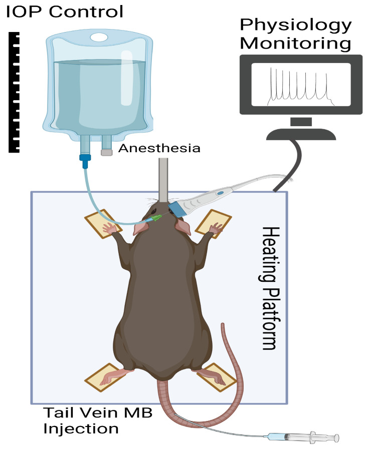
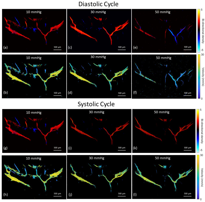
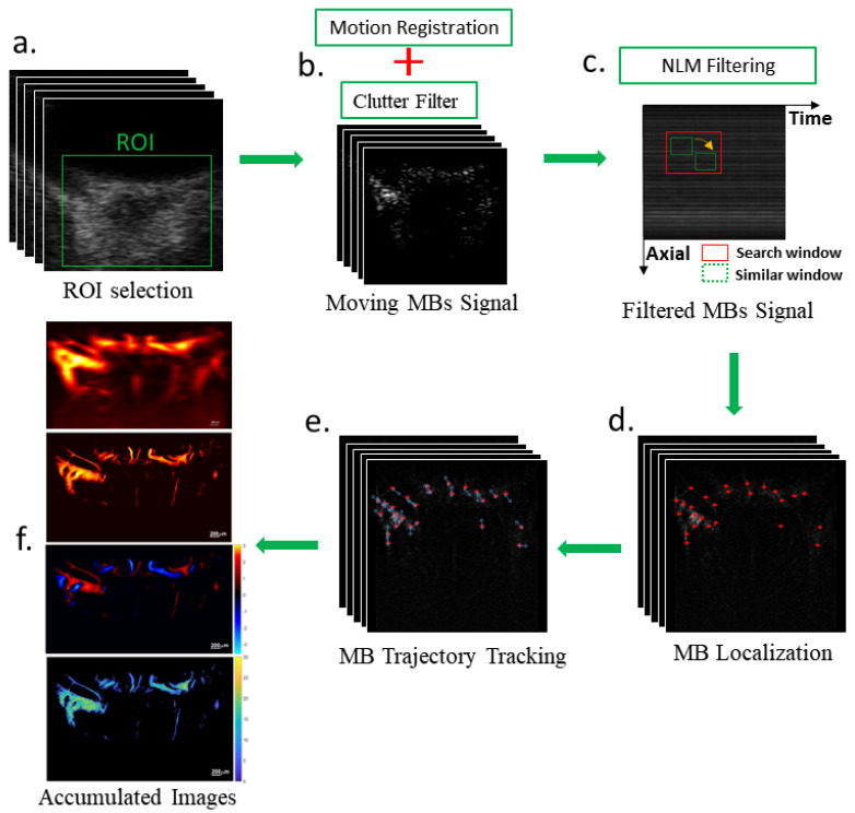
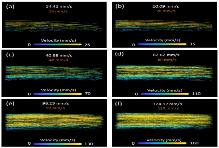
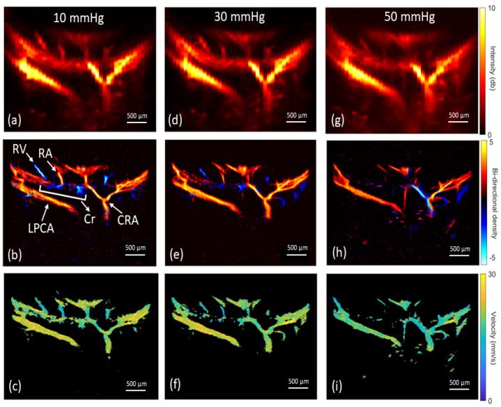
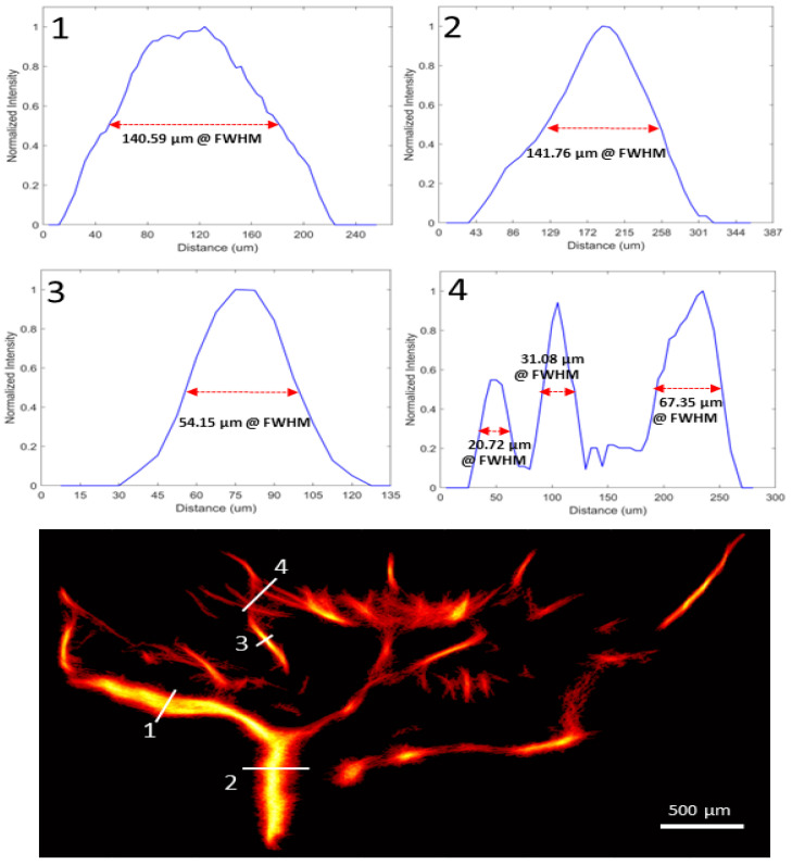
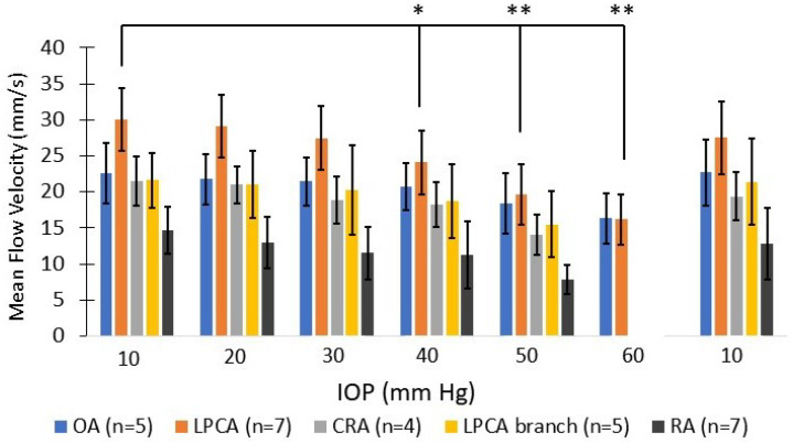
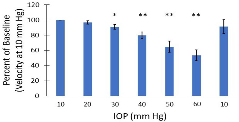
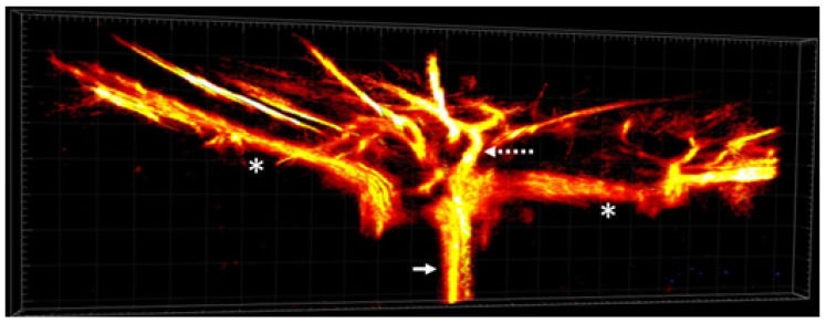
Imaging of the ocular vasculature can provide new insights into the pathophysiology of ocular diseases. This study proposes a novel high-frequency super-resolution ultrasound localization microscopy (SRULM) technique and evaluates its ability to measure in vivo perfusion changes in the rat eye at elevated intraocular pressure (IOP). A 38.4 MHz center frequency linear array transducer on a VisualSonics Vevo F2 imaging platform was used to collect high frame rate (1 kHz) radiofrequency data of the posterior rat eye following systemic microbubble contrast injection. Following clutter and spatiotemporal non-local means filtering, individual microbubbles were localized and tracked. The microbubble tracks were accumulated over 10,000 frames to generate vascular images quantifying perfusion velocity and direction. Experiments were performed using physiologic relevant controlled flow states for algorithm validation and subsequently performed in vivo on the rat eye at 10 mm Hg IOP increments from 10 to 60 mm Hg. The posterior vasculature of the rat eye, including the ophthalmic artery, long posterior ciliary arteries and their branches, central retinal artery and retinal arterioles and venules were successfully visualized, and velocities quantified at each IOP level. Significant reductions in arterial flow were measured as IOP was elevated. High-frequency SRULM can be used to visualize and quantify the perfusion velocity of the rat eye in both the retrobulbar and intraocular vasculature simultaneously. The ability to detect ocular perfusion changes throughout the depth of the eye may help elucidate the role ischemia has in the pathophysiology of ocular diseases such as glaucoma.

摘要

眼部血管成像能够为眼部疾病的病理生理学提供新的见解。本研究提出了一种新型高频超分辨率超声定位显微镜(SRULM)技术,并评估其在眼内压(IOP)升高时测量大鼠眼部体内灌注变化的能力。在VisualSonics Vevo F2成像平台上,使用中心频率为38.4 MHz的线性阵列换能器,在全身注射微泡造影剂后,采集大鼠眼后部的高帧率(1 kHz)射频数据。经过杂波和时空非局部均值滤波后,对单个微泡进行定位和跟踪。微泡轨迹在10000帧上进行累积,以生成量化灌注速度和方向的血管图像。使用生理相关的可控血流状态进行实验以验证算法,随后在大鼠眼上进行体内实验,眼内压以10 mmHg的增量从10 mmHg升高到60 mmHg。成功可视化了大鼠眼的后部血管系统,包括眼动脉、睫状后长动脉及其分支、视网膜中央动脉以及视网膜小动脉和小静脉,并在每个眼内压水平量化了速度。随着眼内压升高,测量到动脉血流显著减少。高频SRULM可用于同时可视化和量化大鼠眼后段和眼内血管系统的灌注速度。在整个眼深度检测眼部灌注变化的能力可能有助于阐明缺血在青光眼等眼部疾病病理生理学中的作用。

https://cdn.ncbi.nlm.nih.gov/pmc/blobs/8e60/10295416/73c117c962d0/bioengineering-10-00689-g010.jpg
https://cdn.ncbi.nlm.nih.gov/pmc/blobs/8e60/10295416/1634c2584fd0/bioengineering-10-00689-g001.jpg
https://cdn.ncbi.nlm.nih.gov/pmc/blobs/8e60/10295416/a53a757bd22c/bioengineering-10-00689-g002.jpg
https://cdn.ncbi.nlm.nih.gov/pmc/blobs/8e60/10295416/bc3df5141a19/bioengineering-10-00689-g003.jpg
https://cdn.ncbi.nlm.nih.gov/pmc/blobs/8e60/10295416/a77d22080d7d/bioengineering-10-00689-g004.jpg
https://cdn.ncbi.nlm.nih.gov/pmc/blobs/8e60/10295416/fc17b3ca1757/bioengineering-10-00689-g005.jpg
https://cdn.ncbi.nlm.nih.gov/pmc/blobs/8e60/10295416/f0c03f2f0b53/bioengineering-10-00689-g006.jpg
https://cdn.ncbi.nlm.nih.gov/pmc/blobs/8e60/10295416/0cbffc64c40e/bioengineering-10-00689-g007.jpg
https://cdn.ncbi.nlm.nih.gov/pmc/blobs/8e60/10295416/b66cee849bf8/bioengineering-10-00689-g008.jpg
https://cdn.ncbi.nlm.nih.gov/pmc/blobs/8e60/10295416/ad281c3aaf4e/bioengineering-10-00689-g009.jpg
https://cdn.ncbi.nlm.nih.gov/pmc/blobs/8e60/10295416/73c117c962d0/bioengineering-10-00689-g010.jpg

相似文献

1
Super-Resolution Ultrasound Localization Microscopy Using High-Frequency Ultrasound to Measure Ocular Perfusion Velocity in the Rat Eye.使用高频超声的超分辨率超声定位显微镜测量大鼠眼部灌注速度
Bioengineering (Basel). 2023 Jun 6;10(6):689. doi: 10.3390/bioengineering10060689.
2
Super-Resolution Ultrasound Localization Microscopy for Visualization of the Ocular Blood Flow.超分辨率超声定位显微镜用于可视化眼部血流。
IEEE Trans Biomed Eng. 2022 May;69(5):1585-1594. doi: 10.1109/TBME.2021.3120368. Epub 2022 Apr 21.
3
Retrobulbar blood flow in rat eyes during acute elevation of intraocular pressure.大鼠眼内压急性升高时眼后的血流。
Exp Eye Res. 2021 Jun;207:108606. doi: 10.1016/j.exer.2021.108606. Epub 2021 Apr 28.
4
In vivo ocular microvasculature imaging in rabbits with 3D ultrasound localization microscopy.在体兔眼微血管成像的 3D 超声定位显微镜。
Ultrasonics. 2023 Aug;133:107022. doi: 10.1016/j.ultras.2023.107022. Epub 2023 May 9.
5
Changes in the retrobulbar arterial circulation after decrease of the elevated intraocular pressure in men and women with primary open angle glaucoma.原发性开角型青光眼患者眼压升高降低后球后动脉循环的变化(男性和女性)
Srp Arh Celok Lek. 2013 Nov-Dec;141(11-12):728-31. doi: 10.2298/sarh1312728m.
6
Super-Resolution Ultrasound Imaging Can Quantify Alterations in Microbubble Velocities in the Renal Vasculature of Rats.超分辨率超声成像可量化大鼠肾血管中微泡速度的变化。
Diagnostics (Basel). 2022 Apr 28;12(5):1111. doi: 10.3390/diagnostics12051111.
7
Effect of intravenous droperidol on intraocular pressure and retrobulbar hemodynamics.静脉注射氟哌利多对眼压和球后血流动力学的影响。
Eur J Ophthalmol. 2002 May-Jun;12(3):193-9. doi: 10.1177/112067210201200304.
8
Impact of intraocular pressure on changes of blood flow in the retina, choroid, and optic nerve head in rats investigated by optical microangiography.通过光学微血管造影术研究眼压对大鼠视网膜、脉络膜和视神经乳头血流变化的影响。
Biomed Opt Express. 2012 Sep 1;3(9):2220-33. doi: 10.1364/BOE.3.002220. Epub 2012 Aug 24.
9
Effects of glaucoma drugs on ocular hemodynamics in normal tension glaucoma: a randomized trial comparing bimatoprost and latanoprost with dorzolamide [ISRCTN18873428].青光眼药物对正常眼压性青光眼患者眼血流动力学的影响:一项比较比马前列素、拉坦前列素与多佐胺的随机试验[国际标准随机对照试验编号:ISRCTN18873428]
BMC Ophthalmol. 2005 Apr 5;5:6. doi: 10.1186/1471-2415-5-6.
10
In Vivo Visualization of Eye Vasculature Using Super-Resolution Ultrasound Microvessel Imaging.利用超分辨率超声微血管成像技术对眼部血管进行体内可视化。
IEEE Trans Biomed Eng. 2020 Oct;67(10):2870-2880. doi: 10.1109/TBME.2020.2972514. Epub 2020 Feb 10.

引用本文的文献

1
Progresses and clinical application of super-resolution ultrasound imaging: a narrative review.超分辨率超声成像的进展与临床应用:一篇叙述性综述
Ultrasound J. 2025 Jun 16;17(1):29. doi: 10.1186/s13089-025-00432-6.
2
Ultrasound super-resolution imaging and shear wave elastography for the non-invasive diagnosis of non-proliferative diabetic retinopathy: a pilot study.超声超分辨率成像与剪切波弹性成像用于非增殖性糖尿病视网膜病变的无创诊断:一项初步研究
Quant Imaging Med Surg. 2025 May 1;15(5):4059-4070. doi: 10.21037/qims-24-1442. Epub 2025 Apr 10.
3
Impact of Elevated Intraocular Pressure on Lamina Cribrosa Oxygenation: A Combined Experimental-Computational Study on Monkeys.

本文引用的文献

1
Retinal functional ultrasound imaging (rfUS) for assessing neurovascular alterations: a pilot study on a rat model of dementia.视网膜功能超声成像(rfUS)评估神经血管改变:痴呆大鼠模型的初步研究。
Sci Rep. 2022 Nov 14;12(1):19515. doi: 10.1038/s41598-022-23366-8.
2
In vivo whole brain microvascular imaging in mice using transcranial 3D Ultrasound Localization Microscopy.在体小鼠全脑微血管成像的颅外 3D 超声定位显微镜技术。
EBioMedicine. 2022 May;79:103995. doi: 10.1016/j.ebiom.2022.103995. Epub 2022 Apr 20.
3
Performance benchmarking of microbubble-localization algorithms for ultrasound localization microscopy.
眼压升高对筛板氧合的影响:一项对猴子的实验与计算相结合的研究
Ophthalmol Sci. 2025 Jan 31;5(3):100725. doi: 10.1016/j.xops.2025.100725. eCollection 2025 May-Jun.
4
Impact of anatomic variability and other vascular factors on lamina cribrosa hypoxia.解剖变异及其他血管因素对筛板缺氧的影响。
bioRxiv. 2024 Sep 13:2024.09.12.610282. doi: 10.1101/2024.09.12.610282.
超声定位显微镜中微泡定位算法的性能基准测试。
Nat Biomed Eng. 2022 May;6(5):605-616. doi: 10.1038/s41551-021-00824-8. Epub 2022 Feb 17.
4
Dynamic Alterations in Blood Flow in Glaucoma Measured with Laser Speckle Contrast Imaging.应用激光散斑对比成像测量青光眼血流的动态变化。
Ophthalmol Glaucoma. 2022 May-Jun;5(3):250-261. doi: 10.1016/j.ogla.2021.10.005. Epub 2021 Oct 18.
5
Super-Resolution Ultrasound Localization Microscopy for Visualization of the Ocular Blood Flow.超分辨率超声定位显微镜用于可视化眼部血流。
IEEE Trans Biomed Eng. 2022 May;69(5):1585-1594. doi: 10.1109/TBME.2021.3120368. Epub 2022 Apr 21.
6
Retinal blood flow reversal quantitatively monitored in out-of-plane vessels with laser Doppler holography.利用激光多普勒全息术定量监测离面血管中的视网膜血流反向。
Sci Rep. 2021 Sep 8;11(1):17828. doi: 10.1038/s41598-021-96877-5.
7
Retrobulbar blood flow in rat eyes during acute elevation of intraocular pressure.大鼠眼内压急性升高时眼后的血流。
Exp Eye Res. 2021 Jun;207:108606. doi: 10.1016/j.exer.2021.108606. Epub 2021 Apr 28.
8
Super-resolution ultrasound localization microscopy based on a high frame-rate clinical ultrasound scanner: an in-human feasibility study.基于高帧率临床超声扫描仪的超分辨率超声定位显微镜:在人体中的可行性研究。
Phys Med Biol. 2021 Apr 8;66(8). doi: 10.1088/1361-6560/abef45.
9
Influence of Choroidal Microvasculature Dropout on the Rate of Glaucomatous Progression: A Prospective Study.脉络膜微血管缺失对青光眼进展速度的影响:一项前瞻性研究。
Ophthalmol Glaucoma. 2020 Jan-Feb;3(1):25-31. doi: 10.1016/j.ogla.2019.10.001. Epub 2019 Oct 25.
10
Progressive retinal nerve fibre layer thinning and choroidal microvasculature dropout at the location of disc haemorrhage in glaucoma.青光眼视盘出血部位视网膜神经纤维层进行性变薄和脉络膜微血管缺失。
Br J Ophthalmol. 2021 May;105(5):674-680. doi: 10.1136/bjophthalmol-2020-316169. Epub 2020 Jul 1.