Department of Small Animal Clinical Sciences, College of Veterinary Medicine, University of Florida, Gainesville, Florida.
Vet Med Sci. 2023 Jul;9(4):1441-1445. doi: 10.1002/vms3.1175. Epub 2023 Jun 29.
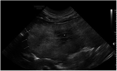
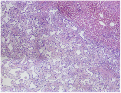
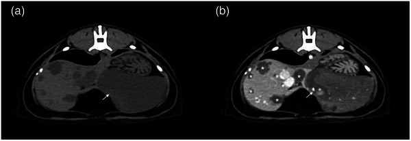
A 13-year-old, male neutered domestic short-haired cat was diagnosed with multiple biliary duct hamartomas after liver lobectomy for a suspected malignant hepatic mass. Distinguishing ultrasonographic findings included a lobular, mostly well-defined, heterogeneous, predominantly hyperechoic, left hepatic mass. Computed tomography (CT) confirmed the presence of a lobular, well-defined, fluid to soft tissue attenuating, heterogeneously hypoenhancing left divisional hepatic mass. Grossly, a large left sided multilobular pale pink gelatinous hepatic mass was surgically excised. Histopathologically, the mass was composed of irregular cystic spaces lined by cuboidal epithelium and separated by mature regular fibrous tissue. Three months following surgery there was no evidence of recurrence or progression of disease on repeat abdominal ultrasound (AUS).
一只 13 岁雄性已绝育的家养短毛猫,因疑似恶性肝肿瘤而行肝叶切除术,术后被诊断为多发性胆管错构瘤。超声表现为边界清楚的分叶状、不均匀的高回声为主的混杂回声团块,位于肝左叶。CT 证实存在边界清楚的分叶状、水样到软组织密度、不均匀低增强的肝左叶肿块。大体检查发现,手术切除了一个大的左侧多小叶状淡粉红色胶冻状肝肿块。组织病理学检查显示,肿块由不规则的囊性腔隙组成,腔隙被立方上皮细胞衬里,由成熟的规则纤维组织分隔。术后 3 个月,重复腹部超声(AUS)未见肿瘤复发或进展。