Conner Sydney, Guarin Justinne R, Le Thanh T, Fatherree Jackson, Kelley Charlotte, Payne Samantha, Salhany Ken, McGinn Rachel, Henrich Emily, Yui Anna, Parker Savannah, Srinivasan Deepti, Bloomer Hanan, Borges Hannah, Oudin Madeleine J
Department of Biomedical Engineering, 200 College Avenue, Tufts University, Medford MA 02155, USA.
bioRxiv. 2023 Jun 14:2023.06.14.544969. doi: 10.1101/2023.06.14.544969.
Metastasis is the leading cause of death in breast cancer patients. For metastasis to occur, tumor cells must invade locally, intravasate, and colonize distant tissues and organs, all steps that require tumor cell migration. The majority of studies on invasion and metastasis rely on human breast cancer cell lines. While it is known that these cells have different properties and abilities for growth and metastasis, the morphological, proliferative, migratory, and invasive behavior of these cell lines and their correlation to behavior is poorly understood. Thus, we sought to classify each cell line as poorly or highly metastatic by characterizing tumor growth and metastasis in a murine model of six commonly used human triple-negative breast cancer xenografts, as well as determine which in vitro assays commonly used to study cell motility best predict metastasis.
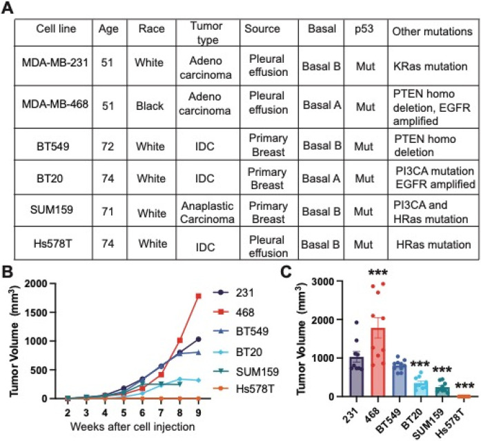
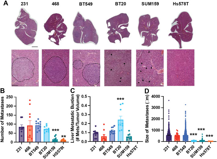
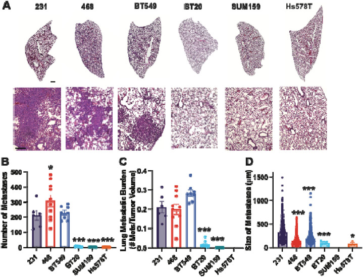
We evaluated the liver and lung metastasis of human TNBC cell lines MDA-MB-231, MDA-MB-468, BT549, Hs578T, BT20, and SUM159 in immunocompromised mice. We characterized each cell line's cell morphology, proliferation, and motility in 2D and 3D to determine the variation in these parameters between cell lines.
We identified MDA-MB-231, MDA-MB-468, and BT549 cells as highly tumorigenic and metastatic, Hs578T as poorly tumorigenic and metastatic, BT20 as intermediate tumorigenic with poor metastasis to the lungs but highly metastatic to the livers, and SUM159 as intermediate tumorigenic but poorly metastatic to the lungs and livers. We showed that metrics that characterize cell morphology are the most predictive of tumor growth and metastatic potential to the lungs and liver. Further, we found that no single motility assay in 2D or 3D significantly correlated with metastasis .
Our results provide an important resource for the TNBC research community, identifying the metastatic potential of 6 commonly used cell lines. Our findings also support the use of cell morphological analysis to investigate the metastatic potential and emphasize the need for multiple motility metrics using multiple cell lines to represent the heterogeneity of metastasis .
转移是乳腺癌患者死亡的主要原因。肿瘤细胞若要发生转移,必须先局部浸润、进入血管,并在远处组织和器官中定植,而这些步骤均需要肿瘤细胞迁移。大多数关于侵袭和转移的研究都依赖于人类乳腺癌细胞系。虽然已知这些细胞在生长和转移方面具有不同的特性和能力,但对这些细胞系的形态、增殖、迁移和侵袭行为及其与转移行为的相关性却知之甚少。因此,我们试图通过在六种常用的人类三阴性乳腺癌异种移植小鼠模型中表征肿瘤生长和转移情况,将每种细胞系分类为低转移或高转移,并确定哪些常用于研究细胞运动性的体外试验最能预测转移。
我们评估了免疫缺陷小鼠中人类三阴性乳腺癌细胞系MDA-MB-231、MDA-MB-468、BT549、Hs578T、BT20和SUM159的肝转移和肺转移情况。我们在二维和三维空间中表征了每种细胞系的细胞形态、增殖和运动性,以确定细胞系之间这些参数的差异。
我们确定MDA-MB-231、MDA-MB-468和BT549细胞具有高致瘤性和转移性,Hs578T致瘤性和转移性较低,BT20致瘤性中等,肺转移较差但肝转移高,SUM159致瘤性中等但肺和肝转移较差。我们表明,表征细胞形态的指标最能预测肿瘤生长以及肺和肝的转移潜力。此外,我们发现二维或三维中的单一运动试验与转移均无显著相关性。
我们的结果为三阴性乳腺癌研究界提供了重要资源,确定了6种常用细胞系的转移潜力。我们的发现还支持使用细胞形态分析来研究转移潜力,并强调需要使用多种细胞系的多个运动指标来代表转移的异质性。