• 文献检索
  • 文档翻译
  • 深度研究
  • 学术资讯
  • Suppr Zotero 插件Zotero 插件
  • 邀请有礼
  • 套餐&价格
  • 历史记录
应用&插件
Suppr Zotero 插件Zotero 插件浏览器插件Mac 客户端Windows 客户端微信小程序
定价
高级版会员购买积分包购买API积分包
服务
文献检索文档翻译深度研究API 文档MCP 服务
关于我们
关于 Suppr公司介绍联系我们用户协议隐私条款
关注我们

Suppr 超能文献

核心技术专利:CN118964589B侵权必究
粤ICP备2023148730 号-1Suppr @ 2026

文献检索

告别复杂PubMed语法,用中文像聊天一样搜索,搜遍4000万医学文献。AI智能推荐,让科研检索更轻松。

立即免费搜索

文件翻译

保留排版,准确专业,支持PDF/Word/PPT等文件格式,支持 12+语言互译。

免费翻译文档

深度研究

AI帮你快速写综述,25分钟生成高质量综述,智能提取关键信息,辅助科研写作。

立即免费体验

基于第一、二磨牙 MRI 分割预测青少年 18 岁以上年龄

Prediction of Age Older than 18 Years in Sub-adults by MRI Segmentation of 1st and 2nd Molars.

机构信息

Institute of Clinical Dentistry, Faculty of Dentistry, University of Oslo, Postboks 1109, Blindern, N-00317, Oslo, Norway.

Department of Forensic Sciences, Oslo University Hospital, Postboks 4950 Nydalen, OUS, Rikshospitalet, 0424, Oslo, Norway.

出版信息

Int J Legal Med. 2023 Sep;137(5):1515-1526. doi: 10.1007/s00414-023-03055-5. Epub 2023 Jul 4.

DOI:10.1007/s00414-023-03055-5
PMID:37402013
原文链接:https://pmc.ncbi.nlm.nih.gov/articles/PMC10421773/
Abstract

PURPOSE

To investigate prediction of age older than 18 years in sub-adults using tooth tissue volumes from MRI segmentation of the entire 1st and 2nd molars, and to establish a model for combining information from two different molars.

MATERIALS AND METHODS

We acquired T2 weighted MRIs of 99 volunteers with a 1.5-T scanner. Segmentation was performed using SliceOmatic (Tomovision©). Linear regression was used to analyse the association between mathematical transformation outcomes of tissue volumes, age, and sex. Performance of different outcomes and tooth combinations were assessed based on the p-value of the age variable, common, or separate for each sex, depending on the selected model. The predictive probability of being older than 18 years was obtained by a Bayesian approach using information from the 1st and 2nd molars both separately and combined.

RESULTS

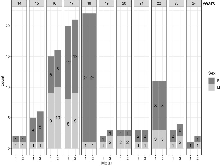
1st molars from 87 participants, and 2nd molars from 93 participants were included. The age range was 14-24 years with a median age of 18 years. The transformation outcome (high signal soft tissue + low signal soft tissue)/total had the strongest statistical association with age for the lower right 1st (p= 7.1*10 for males) and 2nd molar (p=9.44×10 for males and p=7.4×10 for females). Combining the lower right 1st and 2nd molar in males did not increase the prediction performance compared to using the best tooth alone.

CONCLUSION

MRI segmentation of the lower right 1st and 2nd molar might prove useful in the prediction of age older than 18 years in sub-adults. We provided a statistical framework to combine the information from two molars.

摘要

目的

通过对整个第一和第二磨牙的 MRI 分割,研究使用牙组织体积来预测亚成年人年龄超过 18 岁,并建立一种结合来自两个不同磨牙的信息的模型。

材料和方法

我们使用 1.5-T 扫描仪获取了 99 名志愿者的 T2 加权 MRI。使用 SliceOmatic(Tomovision©)进行分割。线性回归用于分析组织体积、年龄和性别之间的数学转换结果之间的关联。基于所选模型,根据年龄变量、共同或单独的 p 值,评估不同结果和牙齿组合的性能。通过使用来自第一和第二磨牙的信息的贝叶斯方法,分别和联合,获得年龄超过 18 岁的预测概率。

结果

纳入了 87 名参与者的第一磨牙和 93 名参与者的第二磨牙。年龄范围为 14-24 岁,中位数年龄为 18 岁。对于右下第一(男性 p=7.1*10)和第二磨牙(男性 p=9.44×10,女性 p=7.4×10),转换结果(高信号软组织+低信号软组织)/总与年龄的统计关联最强。与单独使用最佳牙齿相比,在男性中结合右下第一和第二磨牙并不能提高预测性能。

结论

右下第一和第二磨牙的 MRI 分割可能有助于预测亚成年人年龄超过 18 岁。我们提供了一个统计框架来结合来自两个磨牙的信息。

https://cdn.ncbi.nlm.nih.gov/pmc/blobs/5298/10421773/3cd10d356b90/414_2023_3055_Fig6_HTML.jpg
https://cdn.ncbi.nlm.nih.gov/pmc/blobs/5298/10421773/cfa142ff1c5d/414_2023_3055_Fig1_HTML.jpg
https://cdn.ncbi.nlm.nih.gov/pmc/blobs/5298/10421773/6554ebd57410/414_2023_3055_Fig2_HTML.jpg
https://cdn.ncbi.nlm.nih.gov/pmc/blobs/5298/10421773/04b761d11de1/414_2023_3055_Fig3_HTML.jpg
https://cdn.ncbi.nlm.nih.gov/pmc/blobs/5298/10421773/089f49165cb5/414_2023_3055_Fig4_HTML.jpg
https://cdn.ncbi.nlm.nih.gov/pmc/blobs/5298/10421773/325c2fd19b58/414_2023_3055_Fig5_HTML.jpg
https://cdn.ncbi.nlm.nih.gov/pmc/blobs/5298/10421773/3cd10d356b90/414_2023_3055_Fig6_HTML.jpg

相似文献

1
Prediction of Age Older than 18 Years in Sub-adults by MRI Segmentation of 1st and 2nd Molars.基于第一、二磨牙 MRI 分割预测青少年 18 岁以上年龄
Int J Legal Med. 2023 Sep;137(5):1515-1526. doi: 10.1007/s00414-023-03055-5. Epub 2023 Jul 4.
2
MRI segmentation of tooth tissue in age prediction of sub-adults - a new method for combining data from the 1st, 2nd, and 3rd molars.利用第一、二、三磨牙数据融合的新方法进行未成年个体年龄推断的牙组织 MRI 分割
Int J Legal Med. 2024 May;138(3):939-949. doi: 10.1007/s00414-023-03149-0. Epub 2023 Dec 26.
3
Age prediction in sub-adults based on MRI segmentation of 3rd molar tissue volumes.基于第三磨牙组织体积 MRI 分割的未成年年龄预测。
Int J Legal Med. 2023 May;137(3):753-763. doi: 10.1007/s00414-023-02977-4. Epub 2023 Feb 22.
4
A probability model for assessing age relative to the 18-year old threshold based on magnetic resonance imaging of the knee combined with radiography of third molars in the lower jaw.一种基于膝关节磁共振成像与下颌第三磨牙X线摄影评估相对于18岁阈值的年龄的概率模型。
Forensic Sci Int. 2022 Jan;330:111108. doi: 10.1016/j.forsciint.2021.111108. Epub 2021 Nov 19.
5
The topography of the furcation entrance in Chinese molars. Furcation entrance dimensions.中国磨牙根分叉入口的局部解剖结构。根分叉入口尺寸。
J Clin Periodontol. 1994 Aug;21(7):451-6. doi: 10.1111/j.1600-051x.1994.tb00407.x.
6
Forensic age estimation based on development of third molars: a staging technique for magnetic resonance imaging.基于第三磨牙发育情况的法医年龄估计:一种磁共振成像的分期技术。
J Forensic Odontostomatol. 2017 Dec 1;35(2):117-140.
7
Multi-factorial age estimation: A Bayesian approach combining dental and skeletal magnetic resonance imaging.多因素年龄估计:结合牙和骨骼磁共振成像的贝叶斯方法。
Forensic Sci Int. 2020 Jan;306:110054. doi: 10.1016/j.forsciint.2019.110054. Epub 2019 Nov 15.
8
Prevalence and Pattern of Dental Caries and Their Asssociation with Age and Gender in Preschool Children: An Observational Study.学龄前儿童龋齿的患病率、模式及其与年龄和性别的关联:一项观察性研究
Int J Clin Pediatr Dent. 2020 Sep-Oct;13(5):442-450. doi: 10.5005/jp-journals-10005-1803.
9
Age estimation based on pulp chamber volume of first molars from cone-beam computed tomography images.基于锥形束计算机断层扫描图像中第一磨牙髓腔体积的年龄估计
Forensic Sci Int. 2015 Aug;253:133.e1-7. doi: 10.1016/j.forsciint.2015.05.004. Epub 2015 May 14.
10
Age estimation based on 3D pulp chamber segmentation of first molars from cone-beam-computed tomography by integrated deep learning and level set.基于锥形束 CT 的第一磨牙牙髓腔三维分割的集成深度学习和水平集的年龄估计
Int J Legal Med. 2021 Jan;135(1):365-373. doi: 10.1007/s00414-020-02459-x. Epub 2020 Nov 13.

引用本文的文献

1
Age-related patterns in mandibular third molar eruption: presenting new forensic age assessment reference data from a Chinese population.下颌第三磨牙萌出的年龄相关模式:呈现来自中国人群的新的法医年龄评估参考数据。
Forensic Sci Med Pathol. 2025 Mar;21(1):173-182. doi: 10.1007/s12024-024-00820-9. Epub 2024 May 10.
2
MRI segmentation of tooth tissue in age prediction of sub-adults - a new method for combining data from the 1st, 2nd, and 3rd molars.利用第一、二、三磨牙数据融合的新方法进行未成年个体年龄推断的牙组织 MRI 分割
Int J Legal Med. 2024 May;138(3):939-949. doi: 10.1007/s00414-023-03149-0. Epub 2023 Dec 26.

本文引用的文献

1
Age prediction in sub-adults based on MRI segmentation of 3rd molar tissue volumes.基于第三磨牙组织体积 MRI 分割的未成年年龄预测。
Int J Legal Med. 2023 May;137(3):753-763. doi: 10.1007/s00414-023-02977-4. Epub 2023 Feb 22.
2
Age-dependent decrease in dental pulp cavity volume as a feature for age assessment: a comparative in vitro study using 9.4-T UTE-MRI and CBCT 3D imaging.年龄相关性牙髓腔体积减小作为年龄评估特征的比较性体外研究:使用 9.4-TUTE-MRI 和 CBCT 三维成像。
Int J Legal Med. 2021 Jul;135(4):1599-1609. doi: 10.1007/s00414-021-02603-1. Epub 2021 Apr 26.
3
Volume analysis of second molar pulp chamber using cone beam computed tomography for age estimation in Egyptian adults.
利用锥形束计算机断层扫描对埃及成年人第二磨牙牙髓腔进行容积分析以进行年龄估计。
J Forensic Odontostomatol. 2020 Dec 30;38(3):25-34.
4
Evaluation of secondary dentin formation for forensic age assessment by means of semi-automatic segmented ultrahigh field 9.4 T UTE MRI datasets.利用半自动分割超高场 9.4TUTE MRI 数据集评估法医年龄评估中的二次牙本质形成。
Int J Legal Med. 2020 Nov;134(6):2283-2288. doi: 10.1007/s00414-020-02425-7. Epub 2020 Sep 17.
5
The relevance of body mass index in forensic age assessment of living individuals: an age-adjusted linear regression analysis using multivariable fractional polynomials.体重指数在活体法医年龄评估中的相关性:使用多变量分数多项式进行年龄调整的线性回归分析。
Int J Legal Med. 2020 Sep;134(5):1861-1868. doi: 10.1007/s00414-020-02381-2. Epub 2020 Jul 23.
6
Automatic Age Estimation and Majority Age Classification From Multi-Factorial MRI Data.基于多因素 MRI 数据的自动年龄估计和多数年龄分类。
IEEE J Biomed Health Inform. 2019 Jul;23(4):1392-1403. doi: 10.1109/JBHI.2018.2869606. Epub 2018 Sep 24.
7
Bone age for chronological age determination - statement of the European Society of Paediatric Radiology musculoskeletal task force group.骨龄用于确定实际年龄 - 欧洲儿科放射学会肌肉骨骼工作组的声明。
Pediatr Radiol. 2019 Jun;49(7):979-982. doi: 10.1007/s00247-019-04379-4. Epub 2019 Mar 26.
8
BioAlder: a tool for assessing chronological age based on two radiological methods.BioAlder:一种基于两种放射学方法评估年龄的工具。
Int J Legal Med. 2019 Jul;133(4):1177-1189. doi: 10.1007/s00414-018-1959-5. Epub 2018 Nov 1.
9
Advancing estimation of chronological age by utilizing available evidence based on two radiographical methods.利用基于两种放射学方法的现有证据推进实足年龄估计。
Int J Legal Med. 2019 Jan;133(1):217-229. doi: 10.1007/s00414-018-1848-y. Epub 2018 May 7.
10
A radiographic study of the mandibular third molar root development in different ethnic groups.不同种族下颌第三磨牙牙根发育的影像学研究。
J Forensic Odontostomatol. 2017 Dec 1;35(2):97-108.