Hanai Sho, Yanaka Kiyoyuki, Aiyama Hitoshi, Kajita Michihide, Ishikawa Eiichi
Department of Neurosurgery, Tsukuba Memorial Hospital, Tsukuba, Japan.
Department of Neurosurgery, University of Tsukuba, Tsukuba, Japan.
Surg Neurol Int. 2023 Jun 30;14:224. doi: 10.25259/SNI_279_2023. eCollection 2023.
Intracranial arachnoid cysts (ACs) are developmental anomalies usually filled with cerebrospinal fluid (CSF), rarely resolving throughout life. Here, we present a case of an AC with intracystic hemorrhage and subdural hematoma (SDH) that developed after a minor head injury before gradually disappearing. Neuroimaging demonstrated specific changes from hematoma formation to AC disappearance over time. The mechanisms of this condition are discussed based on imaging data.
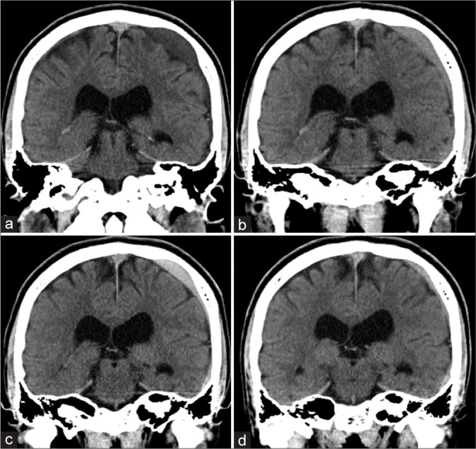
An 18-year-old man was admitted to our hospital with a head injury caused by a traffic accident. On arrival, he was conscious with a mild headache. Computed tomography (CT) revealed no intracranial hemorrhages or skull fractures but an AC was seen in the left convexity. One month later, follow-up CT scans showed an intracystic hemorrhage. Subsequently, an SDH appeared then both the intracystic hemorrhage and SDH gradually shrank, with the AC disappearing spontaneously. The AC was considered to have disappeared, along with the spontaneous SDH resorption.
We present a rare case where neuroimaging demonstrated spontaneous resorption of an AC combined with intracystic hemorrhage and SDH over time, which may provide new insights into the nature of adult ACs.
颅内蛛网膜囊肿(ACs)是一种发育异常,通常充满脑脊液(CSF),很少在一生中自行消退。在此,我们报告一例颅内蛛网膜囊肿合并囊内出血和硬膜下血肿(SDH)的病例,该病例在轻微头部受伤后出现,随后逐渐消失。神经影像学显示了随着时间推移从血肿形成到蛛网膜囊肿消失的特定变化。基于影像学数据对这种情况的机制进行了讨论。
一名18岁男性因交通事故导致头部受伤入院。入院时,他意识清醒,伴有轻度头痛。计算机断层扫描(CT)显示无颅内出血或颅骨骨折,但在左侧脑凸面发现一个蛛网膜囊肿。一个月后,随访CT扫描显示囊内出血。随后,出现硬膜下血肿,随后囊内出血和硬膜下血肿均逐渐缩小,蛛网膜囊肿自发消失。随着硬膜下血肿的自发吸收,蛛网膜囊肿被认为已经消失。
我们报告了一例罕见病例,神经影像学显示颅内蛛网膜囊肿合并囊内出血和硬膜下血肿随时间自发吸收,这可能为成人蛛网膜囊肿的本质提供新的见解。