State Key Laboratory of Oral & Maxillofacial Reconstruction and Regeneration, Key Laboratory of Oral Biomedicine Ministry of Education, Hubei Key Laboratory of Stomatology, School & Hospital of Stomatology, Wuhan University, Wuhan, China.
Department of Oral & Maxillofacial - Head Neck Oncology, School & Hospital of Stomatology, Wuhan University, Wuhan, China.
BMC Oral Health. 2023 Jul 6;23(1):454. doi: 10.1186/s12903-023-03175-9.
Odontogenic keratocyst (OKC) is a relatively common odontogenic lesion characterized by local invasion in the maxillary and mandibular bones. In the pathological tissue slices of OKC, immune cell infiltrations are frequently observed. However, the immune cell profile and the molecular mechanism for immune cell infiltration of OKC are still unclear. We aimed to explore the immune cell profile of OKC and to explore the potential pathogenesis for immune cell infiltration in OKC.
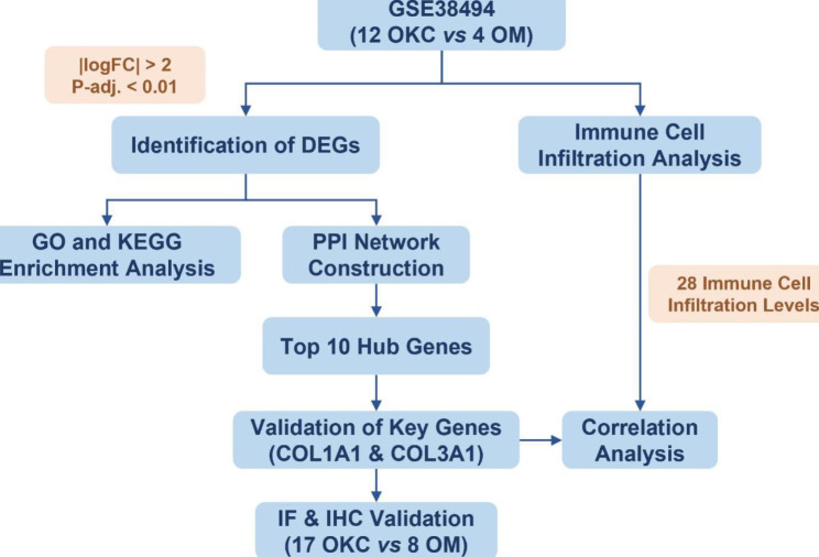
The microarray dataset GSE38494 including OKC and oral mucosa (OM) samples were obtained from the Gene Expression Omnibus (GEO) database. The differentially expressed genes (DEGs) in OKC were analyzed by R software. The hub genes of OKC were performed by protein-protein interaction (PPI) network. The differential immune cell infiltration and the potential relationship between immune cell infiltration and the hub genes were performed by single-sample gene set enrichment analysis (ssGSEA). The expression of COL1A1 and COL1A3 were confirmed by immunofluorescence and immunohistochemistry in 17 OKC and 8 OM samples.
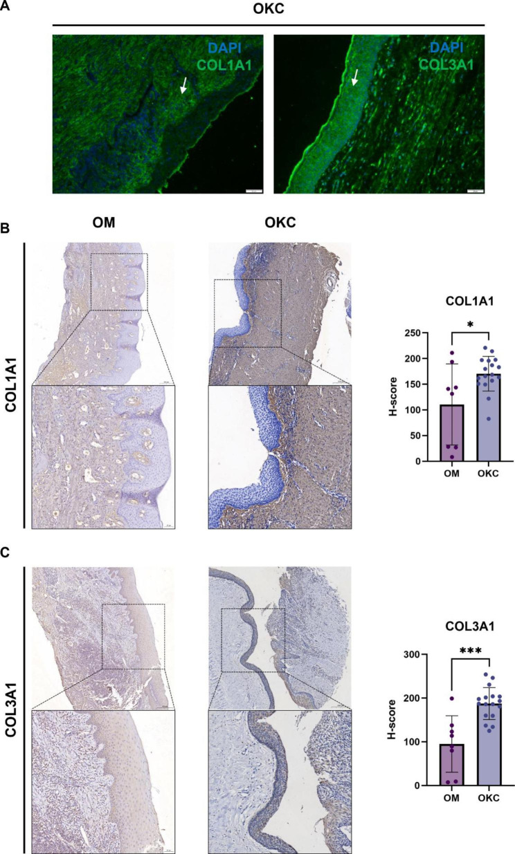
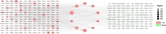
We detected a total of 402 differentially expressed genes (DEGs), of which 247 were upregulated and 155 were downregulated. DEGs were mainly involved in collagen-containing extracellular matrix pathways, external encapsulating structure organization, and extracellular structure organization. We identified ten hub genes, namely FN1, COL1A1, COL3A1, COL1A2, BGN, POSTN, SPARC, FBN1, COL5A1, and COL5A2. A significant difference was observed in the abundances of eight types of infiltrating immune cells between the OM and OKC groups. Both COL1A1 and COL3A1 exhibited a significant positive correlation with natural killer T cells and memory B cells. Simultaneously, they demonstrated a significant negative correlation with CD56dim natural killer cells, neutrophils, immature dendritic cells, and activated dendritic cells. Immunohistochemistry analysis showed that COL1A1 (P = 0.0131) and COL1A3 (P < 0.001) were significantly elevated in OKC compared with OM.
Our findings provide insights into the pathogenesis of OKC and illuminate the immune microenvironment within these lesions. The key genes, including COL1A1 and COL1A3, may significantly impact the biological processes associated with OKC.
牙源性角化囊肿(OKC)是一种较为常见的牙源性病变,其特征为上颌骨和下颌骨局部侵袭。在 OKC 的病理组织切片中,常观察到免疫细胞浸润。然而,OKC 中的免疫细胞特征以及免疫细胞浸润的分子机制尚不清楚。本研究旨在探讨 OKC 的免疫细胞特征,并探索 OKC 中免疫细胞浸润的潜在发病机制。
从基因表达综合数据库(GEO)中获取包含 OKC 和口腔黏膜(OM)样本的微阵列数据集 GSE38494。使用 R 软件分析 OKC 中的差异表达基因(DEGs)。通过蛋白质-蛋白质相互作用(PPI)网络筛选 OKC 的关键基因。通过单样本基因集富集分析(ssGSEA)分析差异免疫细胞浸润和免疫细胞浸润与关键基因之间的潜在关系。通过免疫荧光和免疫组织化学在 17 个 OKC 和 8 个 OM 样本中验证 COL1A1 和 COL1A3 的表达。
共检测到 402 个差异表达基因(DEGs),其中 247 个上调,155 个下调。DEGs 主要涉及胶原富含细胞外基质途径、外部包膜结构组织和细胞外结构组织。鉴定出 10 个关键基因,即 FN1、COL1A1、COL3A1、COL1A2、BGN、POSTN、SPARC、FBN1、COL5A1 和 COL5A2。OM 和 OKC 组之间 8 种浸润免疫细胞的丰度存在显著差异。COL1A1 和 COL3A1 与自然杀伤 T 细胞和记忆 B 细胞呈显著正相关,与 CD56dim 自然杀伤细胞、中性粒细胞、未成熟树突状细胞和活化树突状细胞呈显著负相关。免疫组织化学分析显示,COL1A1(P=0.0131)和 COL1A3(P<0.001)在 OKC 中的表达明显高于 OM。
本研究结果为 OKC 的发病机制提供了新的见解,并阐明了这些病变中的免疫微环境。关键基因,包括 COL1A1 和 COL1A3,可能显著影响与 OKC 相关的生物学过程。