Fan Zicheng, Wei Xiaoyun, Chen Keke, Wang Ling, Xu Mingen
School of Automation, Hangzhou Dianzi University, Hangzhou 310018, China.
Key Laboratory of Medical Information and 3D Bioprinting of Zhejiang Province, Hangzhou Dianzi University, Hangzhou 310018, China.
Micromachines (Basel). 2023 Apr 19;14(4):878. doi: 10.3390/mi14040878.
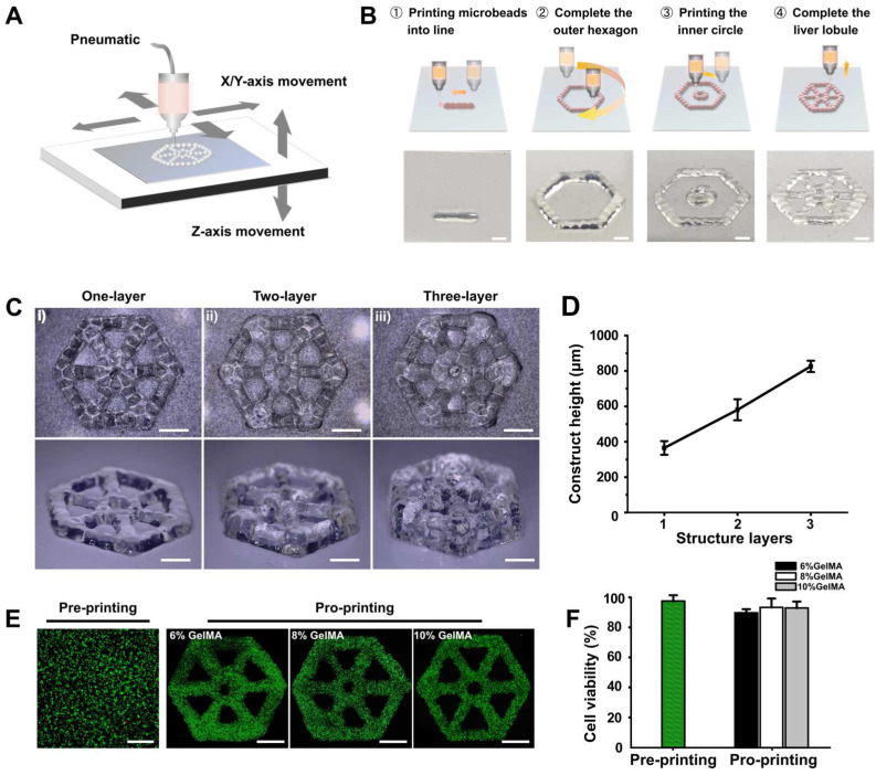
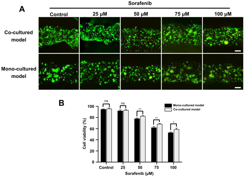
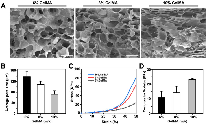
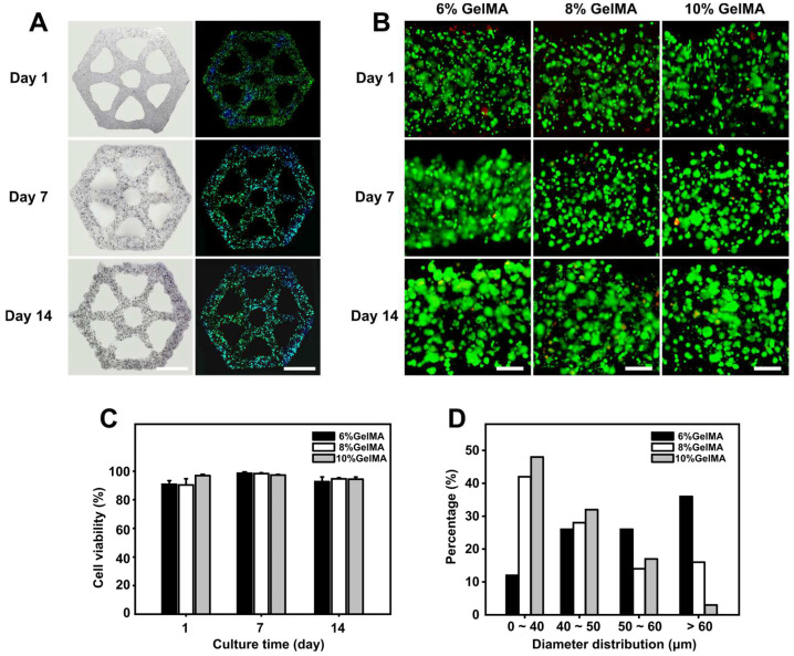
3D cell culture models replicating the complexity of cell-cell interactions and biomimetic extracellular matrix (ECM) are novel approaches for studying liver cancer, including in vitro drug screening or disease mechanism investigation. Although there have been advancements in the production of 3D liver cancer models to serve as drug screening platforms, recreating the structural architecture and tumor-scale microenvironment of native liver tumors remains a challenge. Here, using the dot extrusion printing (DEP) technology reported in our previous work, we fabricated an endothelialized liver lobule-like construct by printing hepatocyte-laden methacryloyl gelatin (GelMA) hydrogel microbeads and HUVEC-laden gelatin microbeads. DEP technology enables hydrogel microbeads to be produced with precise positioning and adjustable scale, facilitating the construction of liver lobule-like structures. The vascular network was achieved by sacrificing the gelatin microbeads at 37 °C to allow HUVEC proliferation on the surface of the hepatocyte layer. Finally, we used the endothelialized liver lobule-like constructs for anti-cancer drug (Sorafenib) screening, and stronger drug resistance results were obtained when compared to either mono-cultured constructs or hepatocyte spheroids alone. The 3D liver cancer models presented here successfully recreate liver lobule-like morphology, and may have the potential to serve as a liver tumor-scale drug screening platform.
复制细胞间相互作用和仿生细胞外基质(ECM)复杂性的3D细胞培养模型是研究肝癌的新方法,包括体外药物筛选或疾病机制研究。尽管在制备用作药物筛选平台的3D肝癌模型方面取得了进展,但重建天然肝肿瘤的结构架构和肿瘤规模的微环境仍然是一项挑战。在这里,我们利用先前工作中报道的点挤出打印(DEP)技术,通过打印载有肝细胞的甲基丙烯酰化明胶(GelMA)水凝胶微珠和载有HUVEC的明胶微珠,制造了一种内皮化的肝小叶样构建体。DEP技术能够精确地定位并以可调节的规模生产水凝胶微珠,有助于构建肝小叶样结构。通过在37°C下牺牲明胶微珠,使HUVEC在肝细胞层表面增殖,从而形成血管网络。最后,我们使用内皮化的肝小叶样构建体进行抗癌药物(索拉非尼)筛选,与单培养构建体或单独的肝细胞球体相比,获得了更强的耐药性结果。本文展示的3D肝癌模型成功地重现了肝小叶样形态,并且可能有潜力作为肝肿瘤规模的药物筛选平台。