• 文献检索
  • 文档翻译
  • 深度研究
  • 学术资讯
  • Suppr Zotero 插件Zotero 插件
  • 邀请有礼
  • 套餐&价格
  • 历史记录
应用&插件
Suppr Zotero 插件Zotero 插件浏览器插件Mac 客户端Windows 客户端微信小程序
定价
高级版会员购买积分包购买API积分包
服务
文献检索文档翻译深度研究API 文档MCP 服务
关于我们
关于 Suppr公司介绍联系我们用户协议隐私条款
关注我们

Suppr 超能文献

核心技术专利:CN118964589B侵权必究
粤ICP备2023148730 号-1Suppr @ 2026

文献检索

告别复杂PubMed语法,用中文像聊天一样搜索,搜遍4000万医学文献。AI智能推荐,让科研检索更轻松。

立即免费搜索

文件翻译

保留排版,准确专业,支持PDF/Word/PPT等文件格式,支持 12+语言互译。

免费翻译文档

深度研究

AI帮你快速写综述,25分钟生成高质量综述,智能提取关键信息,辅助科研写作。

立即免费体验

腹裂和脐膨出胎儿生长过程的多种潜在机制。

Diverse mechanisms underlying the fetal growth course in gastroschisis and omphalocele.

作者信息

Amylidi-Mohr Sofia, Wyss Melanie, Surbek Daniel, Raio Luigi, Mosimann Beatrice

机构信息

Department of Obstetrics, University Hospital of Bern, University of Bern, Bern, Switzerland (Dr Amylidi-Mohr, Ms Wyss, Messrs Surbek and Raio, and Ms Mosimann).

University Hospital of Basel, University of Basel, Basel, Switzerland (Dr Amylidi-Mohr and Ms Mosimann).

出版信息

AJOG Glob Rep. 2023 Jun 4;3(3):100238. doi: 10.1016/j.xagr.2023.100238. eCollection 2023 Aug.

DOI:10.1016/j.xagr.2023.100238
PMID:37424955
原文链接:https://pmc.ncbi.nlm.nih.gov/articles/PMC10329168/
Abstract

BACKGROUND

Gastroschisis and omphalocele are the 2 most common congenital fetal abdominal wall defects. Both malformations are commonly associated with small-for-gestational-age neonates. However, the extent and causes of growth restriction remain controversial in both gastroschisis and omphalocele without associated malformations or aneuploidy.

OBJECTIVE

This study aimed to examine the role of the placenta and the birthweight-to-placental weight ratio in fetuses with abdominal wall defects.

STUDY DESIGN

This study included all cases of abdominal wall defects examined at our hospital between January 2001 and December 2020, retrieving the data from the hospital's software. Fetuses with any other combined congenital anomalies, known chromosomal abnormalities, or lost to follow-up were excluded. Overall, 28 singleton pregnancies with gastroschisis and 24 singleton pregnancies with omphalocele met the inclusion criteria. Patient characteristics and pregnancy outcomes were reviewed. The primary outcome was to investigate the association between birthweight and placental weight in pregnancies with abdominal wall defects as measured after delivery. To correct for gestational age and to compare total placental weights, ratios between the observed and expected birthweights for the given gestational age in singletons were calculated. The scaling exponent β was compared with the reference value of 0.75. Statistical analysis was performed using GraphPad Prism (version 8.2.1; GraphPad Software, San Diego, CA) and IBM SPSS Statistics. A value of <.05 indicated statistical significance.

RESULTS

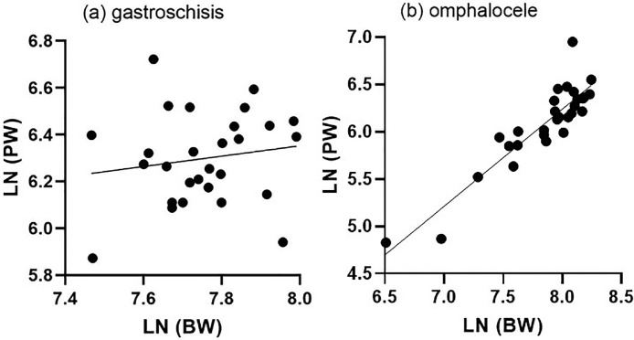
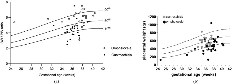
Women pregnant with a fetus with gastroschisis were significantly younger and more often nulliparous. In addition, in this group, the gestational age of delivery was significantly earlier and almost exclusively for cesarean delivery. Of 28 children, 13 (46.7%) were born small for gestational age, only 3 of them (10.7%) had a placental weight <10th percentile. There is no correlation between birthweight percentiles and placental weight percentiles (=not significant). However, in the omphalocele group, 4 of 24 children (16.7%) were born small for gestational age (<10th percentile), and all children also had a placental weight <10th percentile. There is a significant correlation between birthweight percentiles and placental weight percentiles (<.0001). The birthweight-to-placental weight ratio differs significantly between pregnancies diagnosed with gastroschisis and pregnancies diagnosed with omphalocele (4.48 [3.79-4.91] vs 6.05 [5.38-6.47], respectively; <.0001). Allometric metabolic scaling revealed that placentas complicated by gastroschisis and placentas complicated by omphalocele do not scale with birthweight.

CONCLUSION

Fetuses with gastroschisis displayed impaired intrauterine growth, which seemed to differ from the classical placental insufficiency growth restriction.

https://cdn.ncbi.nlm.nih.gov/pmc/blobs/d779/10329168/d02e146b8403/gr4.jpg
https://cdn.ncbi.nlm.nih.gov/pmc/blobs/d779/10329168/67824c8c693a/gr1.jpg
https://cdn.ncbi.nlm.nih.gov/pmc/blobs/d779/10329168/826e8c01cb61/gr2.jpg
https://cdn.ncbi.nlm.nih.gov/pmc/blobs/d779/10329168/fcb35e5f015b/gr3.jpg
https://cdn.ncbi.nlm.nih.gov/pmc/blobs/d779/10329168/d02e146b8403/gr4.jpg
https://cdn.ncbi.nlm.nih.gov/pmc/blobs/d779/10329168/67824c8c693a/gr1.jpg
https://cdn.ncbi.nlm.nih.gov/pmc/blobs/d779/10329168/826e8c01cb61/gr2.jpg
https://cdn.ncbi.nlm.nih.gov/pmc/blobs/d779/10329168/fcb35e5f015b/gr3.jpg
https://cdn.ncbi.nlm.nih.gov/pmc/blobs/d779/10329168/d02e146b8403/gr4.jpg
摘要

背景

腹裂和脐膨出是两种最常见的先天性胎儿腹壁缺陷。这两种畸形通常与小于胎龄儿相关。然而,在无相关畸形或非整倍体的腹裂和脐膨出病例中,生长受限的程度和原因仍存在争议。

目的

本研究旨在探讨胎盘及出生体重与胎盘重量比值在腹壁缺陷胎儿中的作用。

研究设计

本研究纳入了2001年1月至2020年12月在我院检查的所有腹壁缺陷病例,从医院软件中获取数据。排除有任何其他合并先天性异常、已知染色体异常或失访的胎儿。总体而言,28例单胎妊娠合并腹裂和24例单胎妊娠合并脐膨出符合纳入标准。回顾了患者特征和妊娠结局。主要结局是研究分娩后测量的腹壁缺陷妊娠中出生体重与胎盘重量之间的关联。为校正孕周并比较胎盘总重量,计算了单胎中给定孕周的观察出生体重与预期出生体重的比值。将标度指数β与参考值0.75进行比较。使用GraphPad Prism(版本8.2.1;GraphPad软件,加利福尼亚州圣地亚哥)和IBM SPSS Statistics进行统计分析。P值<0.05表示具有统计学意义。

结果

怀有腹裂胎儿的孕妇明显更年轻,且多为初产妇。此外,在该组中,分娩孕周明显更早,且几乎均为剖宫产。28名儿童中,13名(46.7%)出生时为小于胎龄儿,其中只有3名(10.7%)胎盘重量<第10百分位数。出生体重百分位数与胎盘重量百分位数之间无相关性(P =无显著性)。然而,在脐膨出组中,24名儿童中有4名(16.7%)出生时为小于胎龄儿(<第10百分位数),且所有儿童胎盘重量也<第10百分位数。出生体重百分位数与胎盘重量百分位数之间存在显著相关性(P<0.0001)。诊断为腹裂的妊娠与诊断为脐膨出的妊娠之间的出生体重与胎盘重量比值差异显著(分别为4.48[3.79 - 4.91]和6.05[5.38 - 6.47];P<0.0001)。异速生长代谢标度显示,合并腹裂的胎盘和合并脐膨出的胎盘与出生体重不成比例。

结论

腹裂胎儿表现出宫内生长受损,这似乎与经典的胎盘功能不全性生长受限不同。

相似文献

1
Diverse mechanisms underlying the fetal growth course in gastroschisis and omphalocele.腹裂和脐膨出胎儿生长过程的多种潜在机制。
AJOG Glob Rep. 2023 Jun 4;3(3):100238. doi: 10.1016/j.xagr.2023.100238. eCollection 2023 Aug.
2
Maternal serum soluble fms-like tyrosine kinase-1-to-placental growth factor ratio distinguishes growth-restricted from non-growth-restricted small-for-gestational-age fetuses.母体血清可溶性fms样酪氨酸激酶-1与胎盘生长因子的比值可区分生长受限与非生长受限的小于胎龄儿。
AJOG Glob Rep. 2024 Jan 9;4(1):100302. doi: 10.1016/j.xagr.2023.100302. eCollection 2024 Feb.
3
Accuracy of estimated fetal weight assessment in fetuses with abdominal wall defects.腹壁缺陷胎儿估计胎儿体重评估的准确性。
Am J Obstet Gynecol MFM. 2021 Jul;3(4):100385. doi: 10.1016/j.ajogmf.2021.100385. Epub 2021 Apr 23.
4
Omphalocele and gastroschisis: prenatal diagnosis and peripartal management. A case analysis of the years 1989-1997 at the Department of Obstetrics and Gynecology, University of Homburg/Saar.脐膨出与腹裂:产前诊断与围产期管理。对1989年至1997年洪堡/萨尔大学妇产科病例的分析。
Eur J Obstet Gynecol Reprod Biol. 1999 Nov;87(1):47-54. doi: 10.1016/s0301-2115(99)00078-0.
5
Gastroschisis and omphalocele: does either antenatal diagnosis or route of delivery make a difference in perinatal outcome?腹裂和脐膨出:产前诊断或分娩方式会对围产期结局产生影响吗?
Obstet Gynecol. 1990 Aug;76(2):195-9.
6
Characteristics and perinatal course of prenatally diagnosed fetal abdominal wall defects managed in a tertiary center in Japan.日本某三级医疗中心对产前诊断的胎儿腹壁缺损的特征及围产期过程
J Obstet Gynaecol Res. 2009 Feb;35(1):40-7. doi: 10.1111/j.1447-0756.2008.00871.x.
7
Abdominal wall defects and congenital heart disease.腹壁缺损与先天性心脏病。
Ultrasound Obstet Gynecol. 2003 Apr;21(4):334-7. doi: 10.1002/uog.93.
8
Fetal and neonatal outcome in patients with anterior abdominal wall defects (gastroschisis and omphalocele).胎儿和新生儿结局在患有前腹壁缺陷(腹裂和脐膨出)的患者中。
J Perinat Med. 2011 Nov 16;40(1):85-90. doi: 10.1515/JPM.2011.107.
9
Fetal abdominal wall defects in an Australian tertiary setting: contemporary characteristics, ultrasound accuracy, and outcome.澳大利亚三级医疗机构中的胎儿腹壁缺陷:当代特征、超声准确性和结局。
J Matern Fetal Neonatal Med. 2021 Apr;34(8):1269-1276. doi: 10.1080/14767058.2019.1633303. Epub 2019 Jun 26.
10
Interval growth across gestation in pregnancies with fetal gastroschisis.胎儿脐膨出妊娠中孕期隔增长。
Am J Obstet Gynecol MFM. 2021 Sep;3(5):100415. doi: 10.1016/j.ajogmf.2021.100415. Epub 2021 May 31.

引用本文的文献

1
Amniotic fluid: its role in fetal development and beyond.羊水:其在胎儿发育及其他方面的作用。
J Perinatol. 2025 May 8. doi: 10.1038/s41372-025-02313-1.

本文引用的文献

1
Low placental weight and altered metabolic scaling after severe acute respiratory syndrome coronavirus type 2 infection during pregnancy: a prospective multicentric study.严重急性呼吸综合征冠状病毒 2 型感染孕妇胎盘重量低和代谢比例改变的前瞻性多中心研究。
Clin Microbiol Infect. 2022 May;28(5):718-722. doi: 10.1016/j.cmi.2022.02.003. Epub 2022 Feb 10.
2
Placental abnormalities in congenital heart disease.先天性心脏病中的胎盘异常。
Transl Pediatr. 2021 Aug;10(8):2148-2156. doi: 10.21037/tp-20-347.
3
The placenta in fetal growth restriction: What is going wrong?
胎儿生长受限的胎盘:哪里出了问题?
Placenta. 2020 Jul;96:10-18. doi: 10.1016/j.placenta.2020.05.003. Epub 2020 May 11.
4
Editorial: Causes and Consequences of Intrauterine Growth Restriction.社论:宫内生长受限的原因及后果
Front Endocrinol (Lausanne). 2020 Apr 15;11:205. doi: 10.3389/fendo.2020.00205. eCollection 2020.
5
Effects of gestational age at delivery and type of labor on neonatal outcomes among infants with gastroschisis.分娩时的胎龄和分娩方式对先天性腹裂患儿新生儿结局的影响。
J Matern Fetal Neonatal Med. 2021 Jul;34(13):2041-2046. doi: 10.1080/14767058.2019.1656191. Epub 2019 Sep 1.
6
Placental weight and size in relation to fetal growth restriction: a case-control study.胎盘重量和大小与胎儿生长受限的关系:病例对照研究。
J Matern Fetal Neonatal Med. 2021 May;34(9):1356-1360. doi: 10.1080/14767058.2019.1636371. Epub 2019 Jul 3.
7
The Possible Role of Placental Morphometry in the Detection of Fetal Growth Restriction.胎盘形态测量学在胎儿生长受限检测中的可能作用。
Front Physiol. 2019 Jan 8;9:1884. doi: 10.3389/fphys.2018.01884. eCollection 2018.
8
Associations Between the Features of Gross Placental Morphology and Birthweight.胎盘大体形态特征与出生体重之间的关联。
Pediatr Dev Pathol. 2019 May-Jun;22(3):194-204. doi: 10.1177/1093526618789310. Epub 2018 Jul 16.
9
Fetal Medicine Foundation fetal and neonatal population weight charts.胎儿医学基金会胎儿和新生儿人群体重图表。
Ultrasound Obstet Gynecol. 2018 Jul;52(1):44-51. doi: 10.1002/uog.19073. Epub 2018 May 30.
10
Pathophysiology of placental-derived fetal growth restriction.胎盘源性胎儿生长受限的病理生理学。
Am J Obstet Gynecol. 2018 Feb;218(2S):S745-S761. doi: 10.1016/j.ajog.2017.11.577.